1TU4 image
Deposition Date 2004-06-24
Release Date 2004-10-05
Last Version Date 2024-10-16
Entry Detail
PDB ID:
1TU4
Title:
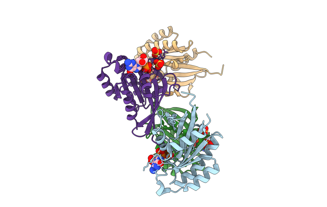
Crystal Structure of Rab5-GDP Complex
Biological Source:
Source Organism:
Homo sapiens (Taxon ID: 9606)
Host Organism:
Method Details:
Experimental Method:
Resolution:
2.20 Å
R-Value Free:
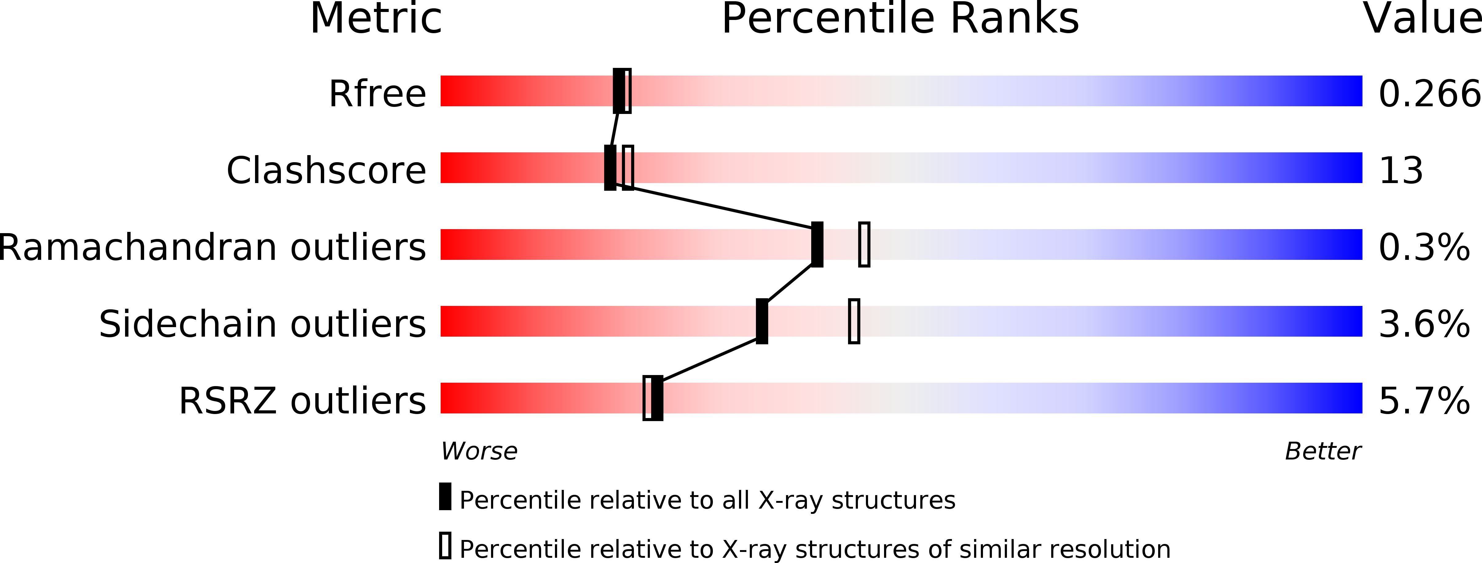
0.27
R-Value Work:
0.21
R-Value Observed:
0.21
Space Group:
P 31 2 1
Macromolecular Entities
Structures with similar UniProt ID
Protein Blast
Polymer Type:polypeptide(L)
Molecule:Ras-related protein Rab-5A
Gene (Uniprot):RAB5A
Chain IDs:A, B, C, D
Chain Length:171
Number of Molecules:4
Biological Source:Homo sapiens
Modified Residue
Compound ID Chain ID Parent Comp ID Details 2D Image
MSE A MET SELENOMETHIONINE
Primary Citation
Structural basis of Rab5-Rabaptin5 interaction in endocytosis
Nat.Struct.Mol.Biol. 11 975 983 (2004)
PMID: 15378032 DOI: 10.1038/nsmb832

Abstact

Rab5 is a small GTPase that regulates early endosome fusion. We present here the crystal structure of the Rab5 GTPase domain in complex with a GTP analog and the C-terminal domain of effector Rabaptin5. The proteins form a dyad-symmetric Rab5-Rabaptin5(2)-Rab5 ternary complex with a parallel coiled-coil Rabaptin5 homodimer in the middle. Two Rab5 molecules bind independently to the Rabaptin5 dimer using their switch and interswitch regions. The binding does not involve the Rab complementarity-determining regions. We also present the crystal structures of two distinct forms of GDP-Rab5 complexes, both of which are incompatible with Rabaptin5 binding. One has a dislocated and disordered switch I but a virtually intact switch II, whereas the other has its beta-sheet and both switch regions reorganized. Biochemical and functional analyses show that the crystallographically observed Rab5-Rabaptin5 complex also exists in solution, and disruption of this complex by mutation abrogates endosome fusion.

Legend

Protein

Chemical

Disease

Primary Citation of related structures