Deposition Date
2004-06-24
Release Date
2004-10-05
Last Version Date
2023-08-23
Entry Detail
PDB ID:
1TU3
Keywords:
Title:
Crystal Structure of Rab5 complex with Rabaptin5 C-terminal Domain
Biological Source:
Source Organism(s):
Homo sapiens (Taxon ID: 9606)
Expression System(s):
Method Details:
Experimental Method:
Resolution:
2.31 Å
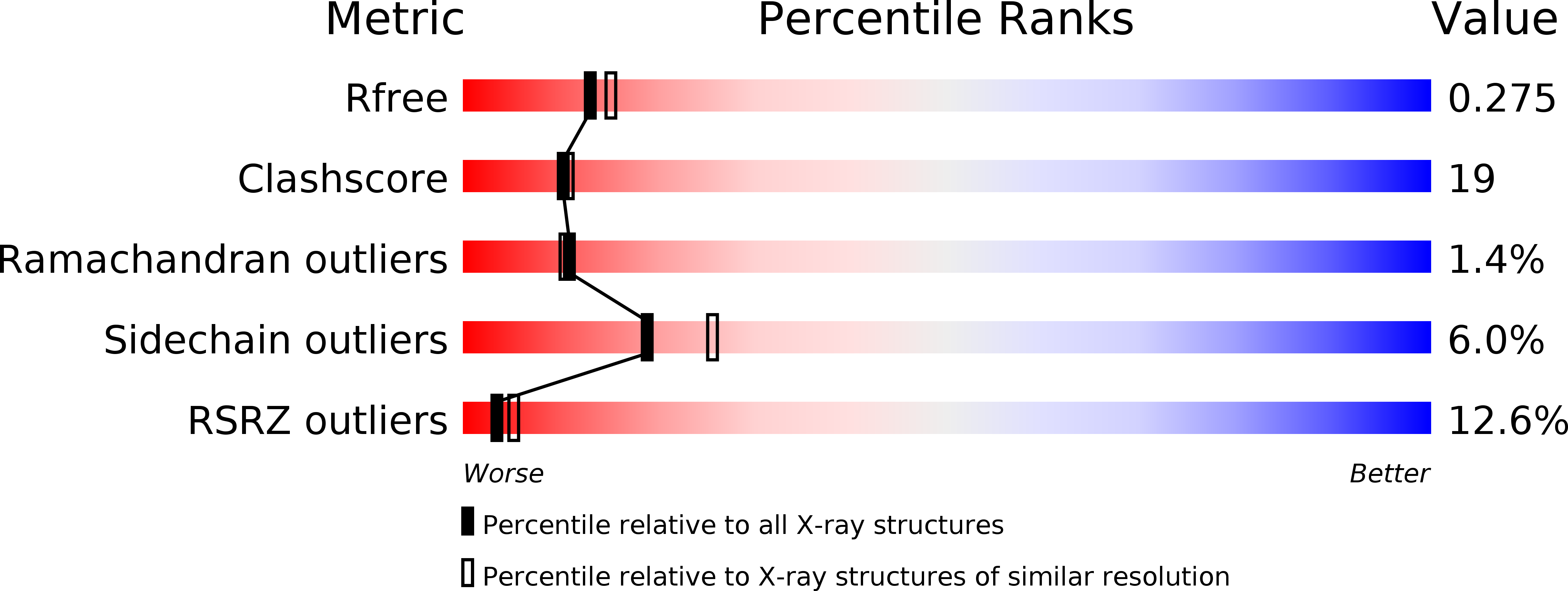
R-Value Free:
0.27
R-Value Work:
0.22
R-Value Observed:
0.22
Space Group:
C 1 2 1