
Deposition Date
1992-11-19
Release Date
1994-01-31
Last Version Date
2024-02-14
Entry Detail
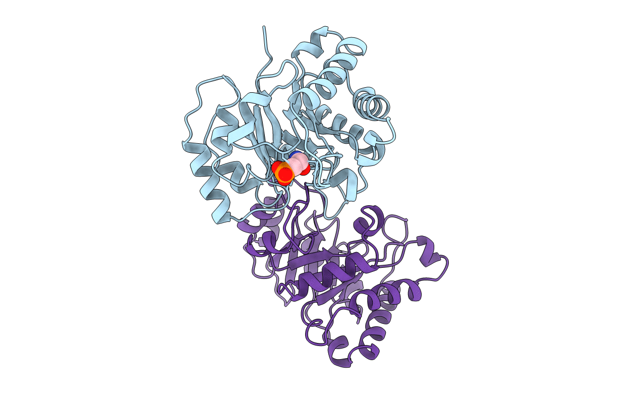
PDB ID:
1TSI
Keywords:
Title:
STRUCTURE OF THE COMPLEX BETWEEN TRYPANOSOMAL TRIOSEPHOSPHATE ISOMERASE AND N-HYDROXY-4-PHOSPHONO-BUTANAMIDE: BINDING AT THE ACTIVE SITE DESPITE AN "OPEN" FLEXIBLE LOOP
Biological Source:
Source Organism(s):
Trypanosoma brucei brucei (Taxon ID: 5702)
Method Details:
Experimental Method:
Resolution:
2.84 Å
R-Value Observed:
0.11
Space Group:
P 21 21 21
