Deposition Date
2004-06-19
Release Date
2004-11-16
Last Version Date
2024-05-22
Entry Detail
PDB ID:
1TR4
Keywords:
Title:
Solution structure of human oncogenic protein gankyrin
Biological Source:
Source Organism(s):
Homo sapiens (Taxon ID: 9606)
Expression System(s):
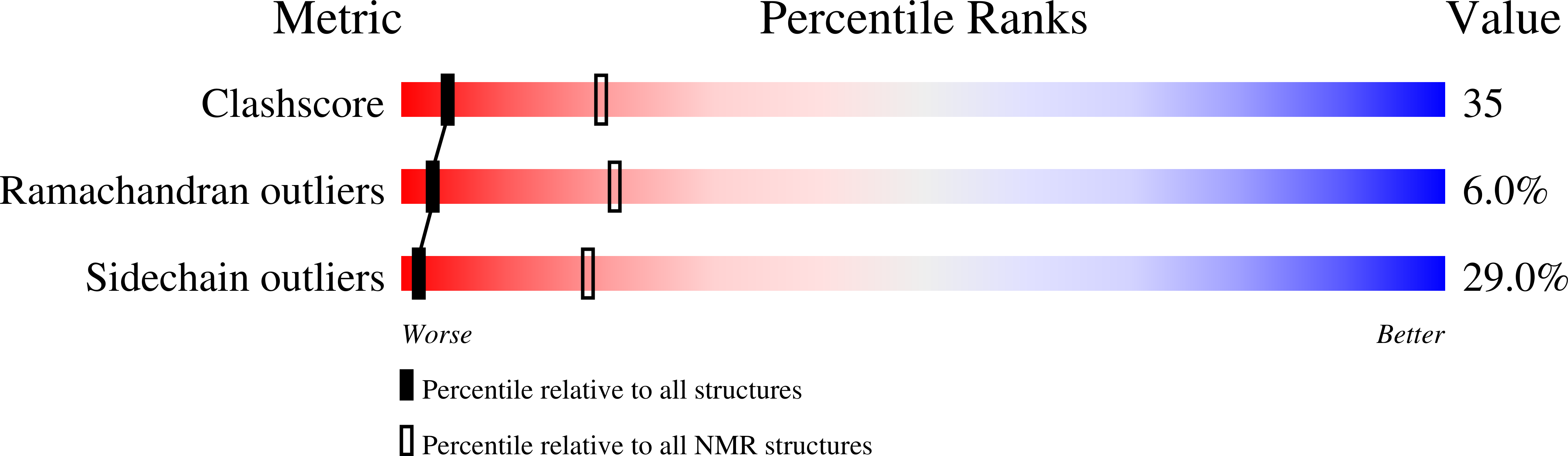
Method Details:
Experimental Method:
Conformers Calculated:
80
Conformers Submitted:
20
Selection Criteria:
structures with the least restraint violations,structures with the lowest energy