1TQJ image
Deposition Date 2004-06-17
Release Date 2004-08-31
Last Version Date 2024-02-14
Entry Detail
PDB ID:
1TQJ
Keywords:
Title:
Crystal structure of D-ribulose 5-phosphate 3-epimerase from Synechocystis to 1.6 angstrom resolution
Biological Source:
Source Organism:
Host Organism:
Method Details:
Experimental Method:
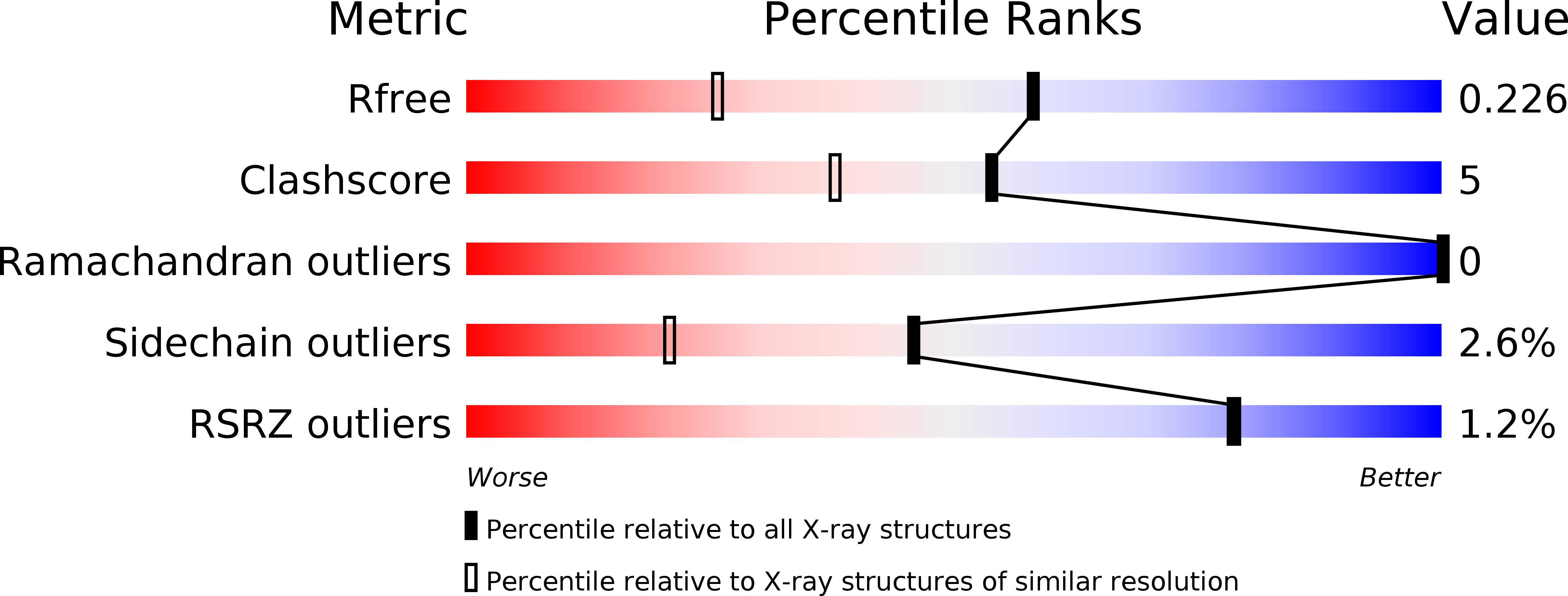
Resolution:
1.60 Å
R-Value Free:
0.21
R-Value Work:
0.17
R-Value Observed:
0.17
Space Group:
P 1 21 1
Macromolecular Entities
Polymer Type:polypeptide(L)
Molecule:Ribulose-phosphate 3-epimerase
Gene (Uniprot):rpe
Chain IDs:A, B, C, D, E, F
Chain Length:230
Number of Molecules:6
Biological Source:Synechocystis sp.
Primary Citation
Structure of D-ribulose 5-phosphate 3-epimerase from Synechocystis to 1.6 A resolution.
Acta Crystallogr.,Sect.D 60 1687 1690 (2004)
PMID: 15333955 DOI: 10.1107/S0907444904015896

Abstact

The crystal structure of D-ribulose 5-phosphate 3-epimerase (RPE) from the cyanobacterium Synechocystis was determined by X-ray crystallography to 1.6 A resolution. The enzyme, which catalyzes the epimerization of D-ribulose 5-phosphate and D-xylulose 5-phosphate, assembles as a hexamer of (beta/alpha)(8)-barrels in the crystallographic asymmetric unit. The active site is highly similar to those of two previously reported RPEs and provides further evidence for essential catalytic roles for several active-site residues.

Legend

Protein

Chemical

Disease

Primary Citation of related structures