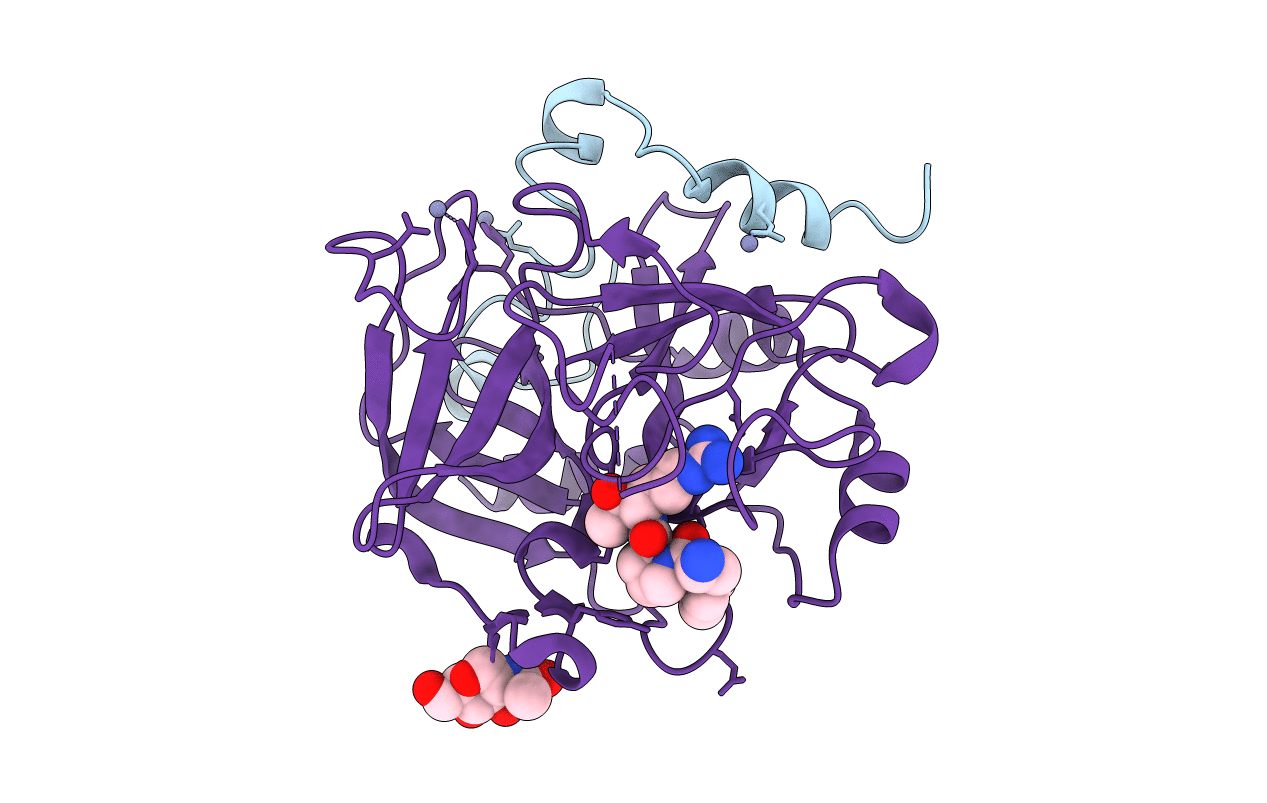
1TQ7 image
Entry Detail
PDB ID:
1TQ7
Title:
Crystal structure of the anticoagulant thrombin mutant W215A/E217A bound to PPACK
Biological Source:
Source Organism:
Host Organism:
PDB Version:
Deposition Date:
2004-06-16
Release Date:
2004-08-03
Method Details:
Experimental Method:
Resolution:
2.40 Å
R-Value Free:
0.24
R-Value Work:
0.20
R-Value Observed:
0.20
Space Group:
P 21 21 21
Macromolecular Entities
Polymer Type:polypeptide(L)
Description:Prothrombin
Chain IDs:A
Chain Length:44
Number of Molecules:1
Biological Source:Homo sapiens
Polymer Type:polypeptide(L)
Description:Prothrombin
Mutations:W215A/E217A
Chain IDs:B
Chain Length:257
Number of Molecules:1
Biological Source:Homo sapiens
Modified Residue
Compound ID Chain ID Parent Comp ID Details 2D Image
ASN B ASN GLYCOSYLATION SITE
Peptide-like Molecules
PRD_000020
Primary Citation
The anticoagulant thrombin mutant W215A/E217A has a collapsed primary specificity pocket
J.Biol.Chem. 279 39824 39828 (2004)
PMID: 15252033 DOI: 10.1074/jbc.M407272200

Abstact

The thrombin mutant W215A/E217A features a drastically impaired catalytic activity toward chromogenic and natural substrates but efficiently activates the anticoagulant protein C in the presence of thrombomodulin. As the remarkable anticoagulant properties of this mutant continue to be unraveled in preclinical studies, we solved the x-ray crystal structures of its free form and its complex with the active site inhibitor H-d-Phe-Pro-Arg-CH(2)Cl (PPACK). The PPACK-bound structure of W215A/E217A is identical to the structure of the PPACK-bound slow form of thrombin. On the other hand, the structure of the free form reveals a collapse of the 215-217 strand that crushes the primary specificity pocket. The collapse results from abrogation of the stacking interaction between Phe-227 and Trp-215 and the polar interactions of Glu-217 with Thr-172 and Lys-224. Other notable changes are a rotation of the carboxylate group of Asp-189, breakage of the H-bond between the catalytic residues Ser-195 and His-57, breakage of the ion pair between Asp-222 and Arg-187, and significant disorder in the 186- and 220-loops that define the Na(+) site. These findings explain the impaired catalytic activity of W215A/E217A and demonstrate that the analysis of the molecular basis of substrate recognition by thrombin and other proteases requires crystallization of both the free and bound forms of the enzyme.

Legend

Protein

Chemical

Disease

Primary Citation of related structures