1TMU image
Deposition Date 1994-05-26
Release Date 1994-09-30
Last Version Date 2024-10-23
Entry Detail
PDB ID:
1TMU
Title:
Changes in interactions in complexes of hirudin derivatives and human alpha-thrombin due to different crystal forms
Biological Source:
Source Organism:
HIRUDO MEDICINALIS (Taxon ID: 6421)
Homo sapiens (Taxon ID: 9606)
Method Details:
Experimental Method:
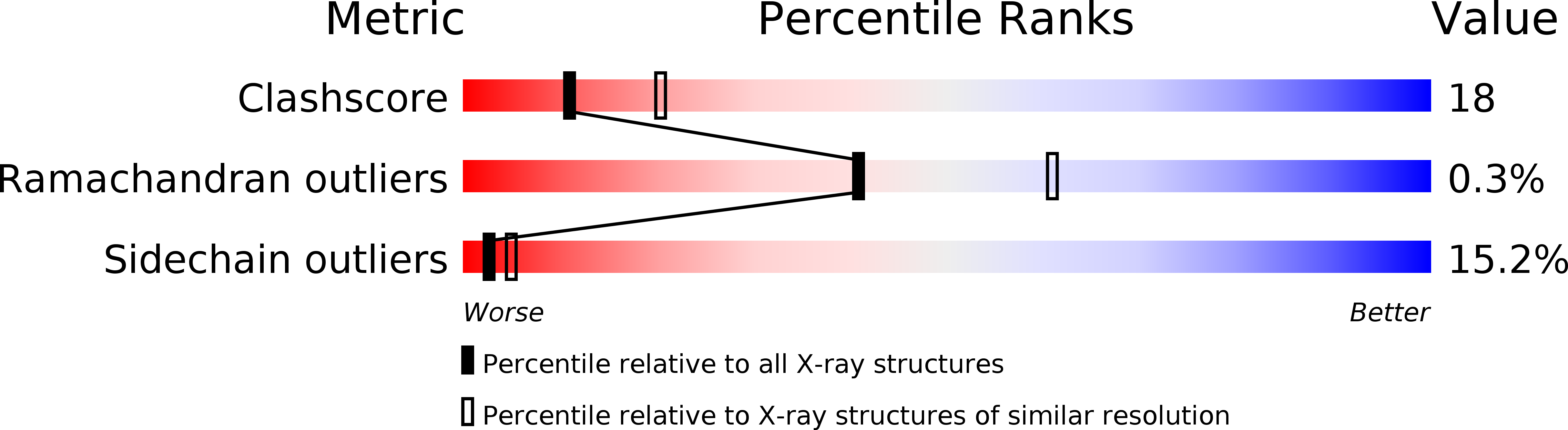
Resolution:
2.50 Å
R-Value Observed:
0.20
Space Group:
P 21 21 2
Macromolecular Entities
Polymer Type:polypeptide(L)
Molecule:Thrombin heavy chain
Gene (Uniprot):F2
Chain IDs:B (auth: H)
Chain Length:259
Number of Molecules:1
Biological Source:Homo sapiens
Polymer Type:polypeptide(L)
Molecule:Hirudin variant-2
Chain IDs:C (auth: J)
Chain Length:11
Number of Molecules:1
Biological Source:HIRUDO MEDICINALIS
Polymer Type:polypeptide(L)
Molecule:Thrombin light chain
Gene (Uniprot):F2
Chain IDs:A (auth: L)
Chain Length:28
Number of Molecules:1
Biological Source:Homo sapiens
Modified Residue
Compound ID Chain ID Parent Comp ID Details 2D Image
ASN B ASN GLYCOSYLATION SITE
TYS C TYR O-SULFO-L-TYROSINE
Peptide-like Molecules
PRD_000020
Primary Citation
Changes in interactions in complexes of hirudin derivatives and human alpha-thrombin due to different crystal forms.
Protein Sci. 2 1630 1642 (1993)
PMID: 8251938

Abstact

The three-dimensional structures of D-Phe-Pro-Arg-chloromethyl ketone-inhibited thrombin in complex with Tyr-63-sulfated hirudin (ternary complex) and of thrombin in complex with the bifunctional inhibitor D-Phe-Pro-Arg-Pro-(Gly)4-hirudin (CGP 50,856, binary complex) have been determined by X-ray crystallography in crystal forms different from those described by Skrzypczak-Jankun et al. (Skrzypczak-Jankun, E., Carperos, V.E., Ravichandran, K.G., & Tulinsky, A., 1991, J. Mol. Biol. 221, 1379-1393). In both complexes, the interactions of the C-terminal hirudin segments of the inhibitors binding to the fibrinogen-binding exosite of thrombin are clearly established, including residues 60-64, which are disordered in the earlier crystal form. The interactions of the sulfate group of Tyr-63 in the ternary complex structure explain why natural sulfated hirudin binds with a 10-fold lower K(i) than the desulfated recombinant material. In this new crystal form, the autolysis loop of thrombin (residues 146-150), which is disordered in the earlier crystal form, is ordered due to crystal contacts. Interactions between the C-terminal fragment of hirudin and thrombin are not influenced by crystal contacts in this new crystal form, in contrast to the earlier form. In the bifunctional inhibitor-thrombin complex, the peptide bond between Arg-Pro (P1-P1') seems to be cleaved.

Legend

Protein

Chemical

Disease

Primary Citation of related structures