Deposition Date
1998-08-03
Release Date
1999-03-30
Last Version Date
2024-02-14
Entry Detail
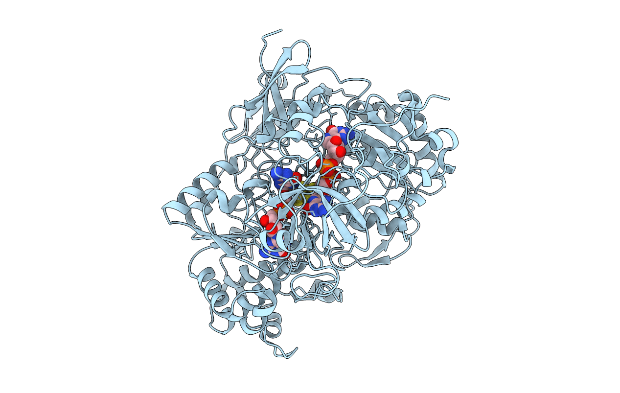
PDB ID:
1TMO
Keywords:
Title:
TRIMETHYLAMINE N-OXIDE REDUCTASE FROM SHEWANELLA MASSILIA
Biological Source:
Source Organism:
Shewanella massilia (Taxon ID: 76854)
Method Details:
Experimental Method:
Resolution:
2.50 Å
R-Value Free:
0.24
R-Value Work:
0.18
R-Value Observed:
0.18
Space Group:
P 21 21 2