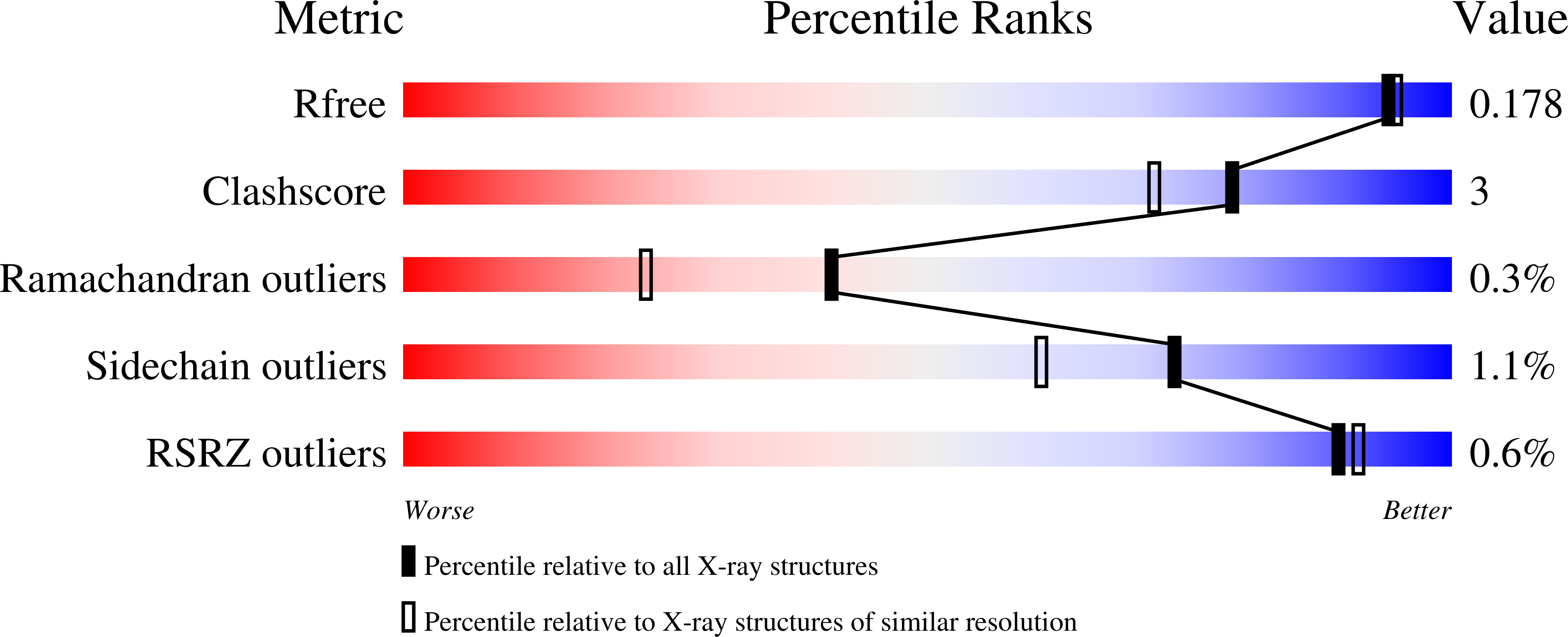
1TMG image
Deposition Date 2004-06-10
Release Date 2004-11-09
Last Version Date 2023-08-23
Entry Detail
PDB ID:
1TMG
Keywords:
Title:
crystal structure of the complex of subtilisin BPN' with chymotrypsin inhibitor 2 M59F mutant
Biological Source:
Host Organism:
Method Details:
Experimental Method:
Resolution:
1.67 Å
R-Value Free:
0.17
R-Value Work:
0.14
R-Value Observed:
0.15
Space Group:
P 65 2 2
Macromolecular Entities
Polymer Type:polypeptide(L)
Molecule:Subtilisin BPN'
Gene (Uniprot):apr
Mutations:C-terminal, 6-His tag
Chain IDs:A (auth: E)
Chain Length:281
Number of Molecules:1
Biological Source:Bacillus amyloliquefaciens
Polymer Type:polypeptide(L)
Molecule:chymotrypsin inhibitor 2
Gene (Uniprot):Ica-2
Mutations:I20, initiating Met; I45A, I59F
Chain IDs:B (auth: I)
Chain Length:64
Number of Molecules:1
Biological Source:Hordeum vulgare subsp. vulgare
Primary Citation
Binding, Proteolytic, and Crystallographic Analyses of Mutations at the Protease-Inhibitor Interface of the Subtilisin BPN'/Chymotrypsin Inhibitor 2 Complex(,).
Biochemistry 43 13648 13656 (2004)
PMID: 15504027 DOI: 10.1021/bi048797k

Abstact

A series of mutants of chymotrypsin inhibitor 2 (CI2), at residues that interact with the inhibited enzyme subtilisin BPN', were studied to determine the relative importance of intermolecular contacts on either side of the scissile bond. Mutants were tested for inhibition of subtilisin, rates of hydrolysis by subtilisin, and ability to acylate subtilisin. Additionally, crystal structures of the mutant CI2 complexes with subtilisin were obtained. Ordered water molecules were found to play an important role in inhibitor recognition, and features of the crystal structures, in combination with biochemical data, support a transition-state stabilization role for the P(1) residue in subtilisin catalysis. Consistent with the proposed mechanism of inhibition, in which rapid acylation is followed by religation, leaving-group contacts with the enzyme were found to be more critical determinants of inhibition than acylating-group contacts in the mutants studied here.

Legend

Protein

Chemical

Disease

Primary Citation of related structures