1TLV image
Deposition Date 2004-06-10
Release Date 2005-02-08
Last Version Date 2024-11-13
Entry Detail
PDB ID:
1TLV
Keywords:
Title:
Structure of the native and inactive LicT PRD from B. subtilis
Biological Source:
Source Organism(s):
Expression System(s):
Method Details:
Experimental Method:
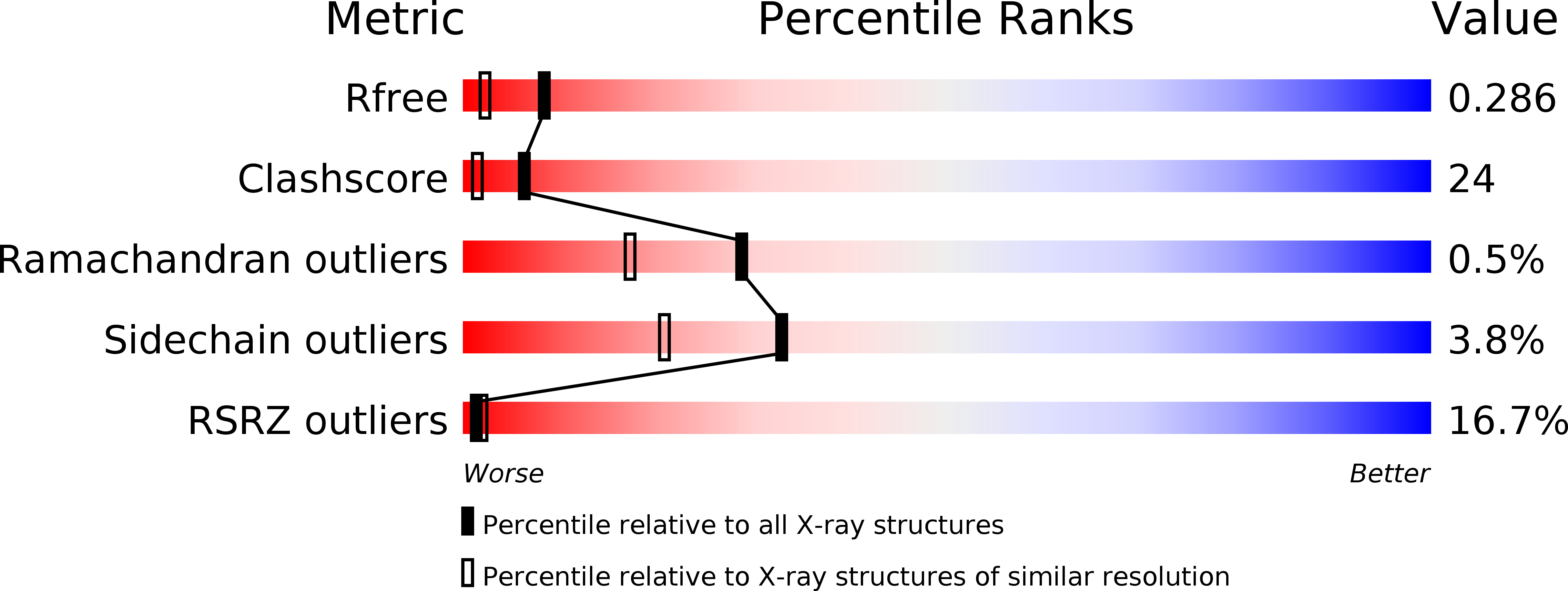
Resolution:
1.95 Å
R-Value Free:
0.29
R-Value Work:
0.26
R-Value Observed:
0.26
Space Group:
P 32 2 1
Macromolecular Entities
Structures with similar UniProt ID
Protein Blast
Polymer Type:polypeptide(L)
Molecule:Transcription antiterminator licT
Gene (Uniprot):licT
Chain IDs:A
Chain Length:221
Number of Molecules:1
Biological Source:Bacillus subtilis
Modified Residue
Compound ID Chain ID Parent Comp ID Details 2D Image
CME A CYS S,S-(2-HYDROXYETHYL)THIOCYSTEINE
Primary Citation
Activation of the LicT Transcriptional Antiterminator Involves a Domain Swing/Lock Mechanism Provoking Massive Structural Changes
J.Biol.Chem. 280 14780 14789 (2005)
PMID: 15699035 DOI: 10.1074/jbc.M414642200

Abstact

The transcriptional antiterminator protein LicT regulates the expression of Bacillus subtilis operons involved in beta-glucoside metabolism. It consists of an N-terminal RNA-binding domain (co-antiterminator (CAT)) and two phosphorylatable phosphotransferase system regulation domains (PRD1 and PRD2). In the activated state, each PRD forms a dimeric unit with the phosphorylation sites totally buried at the dimer interface. Here we present the 1.95 A resolution structure of the inactive LicT PRDs as well as the molecular solution structure of the full-length protein deduced from small angle x-ray scattering. Comparison of native (inactive) and mutant (constitutively active) PRD crystal structures shows massive tertiary and quaternary rearrangements of the entire regulatory domain. In the inactive state, a wide swing movement of PRD2 results in dimer opening and brings the phosphorylation sites to the protein surface. This movement is accompanied by additional structural rearrangements of both the PRD1-PRD1 ' interface and the CAT-PRD1 linker. Small angle x-ray scattering experiments indicate that the amplitude of the PRD2 swing might even be wider in solution than in the crystals. Our results suggest that PRD2 is highly mobile in the native protein, whereas it is locked upon activation by phosphorylation.

Legend

Protein

Chemical

Disease

Primary Citation of related structures
Feedback Form
Name
Email
Institute
Feedback