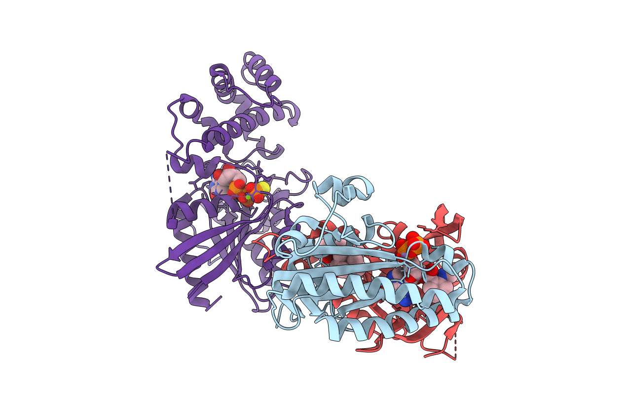
1TL7 image
Deposition Date 2004-06-09
Release Date 2004-12-14
Last Version Date 2023-08-23
Entry Detail
PDB ID:
1TL7
Keywords:
Title:
Complex Of Gs- With The Catalytic Domains Of Mammalian Adenylyl Cyclase: Complex With 2'(3')-O-(N-methylanthraniloyl)-guanosine 5'-triphosphate and Mn
Biological Source:
Source Organism:
Host Organism:
Method Details:
Experimental Method:
Resolution:
2.80 Å
R-Value Free:
0.29
R-Value Work:
0.25
R-Value Observed:
0.25
Space Group:
P 21 21 2
Macromolecular Entities
Polymer Type:polypeptide(L)
Molecule:Adenylate cyclase, type V
Gene (Uniprot):ADCY5
Chain IDs:A
Chain Length:217
Number of Molecules:1
Biological Source:Canis lupus familiaris
Polymer Type:polypeptide(L)
Molecule:Adenylate cyclase, type II
Gene (Uniprot):Adcy2
Chain IDs:B
Chain Length:212
Number of Molecules:1
Biological Source:Rattus norvegicus
Polymer Type:polypeptide(L)
Molecule:Guanine nucleotide-binding protein G(s), alpha subunit
Gene (Uniprot):GNAS
Chain IDs:C
Chain Length:402
Number of Molecules:1
Biological Source:Bos taurus
Primary Citation
Structural basis for the inhibition of mammalian membrane adenylyl cyclase by 2 '(3')-O-(N-Methylanthraniloyl)-guanosine 5 '-triphosphate.
J.Biol.Chem. 280 7253 7261 (2005)
PMID: 15591060 DOI: 10.1074/jbc.M409076200

Abstact

Membrane-bound mammalian adenylyl cyclase (mAC) catalyzes the synthesis of intracellular cyclic AMP from ATP and is activated by stimulatory G protein alpha subunits (Galpha(s)) and by forskolin (FSK). mACs are inhibited with high potency by 2 '(3')-O-(N-methylanthraniloyl) (MANT)-substituted nucleotides. In this study, the crystal structures of the complex between Galpha(s).GTPgammaS and the catalytic C1 and C2 domains from type V and type II mAC (VC1.IIC2), bound to FSK and either MANT-GTP.Mg(2+) or MANT-GTP.Mn(2+) have been determined. MANT-GTP coordinates two metal ions and occupies the same position in the catalytic site as P-site inhibitors and substrate analogs. However, the orientation of the guanine ring is reversed relative to that of the adenine ring. The MANT fluorophore resides in a hydrophobic pocket at the interface between the VC1 and IIC2 domains and prevents mAC from undergoing the "open" to "closed" domain rearrangement. The K(i) of MANT-GTP for inhibition of VC1.IIC2 is lower in the presence of mAC activators and lower in the presence of Mn(2+) compared with Mg(2+), indicating that the inhibitor binds more tightly to the catalytically most active form of the enzyme. Fluorescence resonance energy transfer-stimulated emission from the MANT fluorophore upon excitation of Trp-1020 in the MANT-binding pocket of IIC2 is also stronger in the presence of FSK. Mutational analysis of two non-conserved amino acids in the MANT-binding pocket suggests that residues outside of the binding site influence isoform selectivity toward MANT-GTP.

Legend

Protein

Chemical

Disease

Primary Citation of related structures