1TIW image
Deposition Date 2004-06-02
Release Date 2004-10-26
Last Version Date 2023-08-23
Entry Detail
PDB ID:
1TIW
Keywords:
Title:
Crystal structure of E. coli PutA proline dehydrogenase domain (residues 86-669) complexed with L-Tetrahydro-2-furoic acid
Biological Source:
Source Organism:
Escherichia coli (Taxon ID: 562)
Host Organism:
Method Details:
Experimental Method:
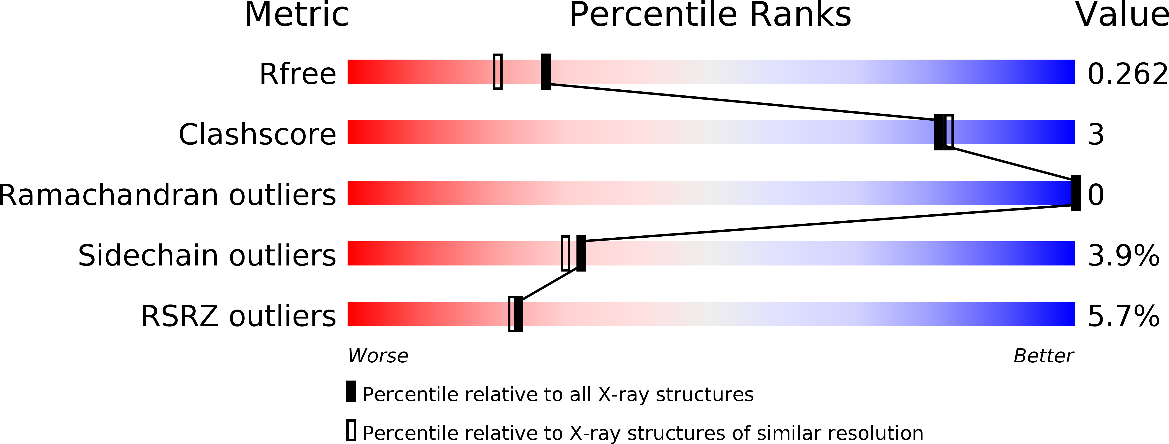
Resolution:
2.00 Å
R-Value Free:
0.25
R-Value Work:
0.21
R-Value Observed:
0.21
Space Group:
I 2 2 2
Macromolecular Entities
Structures with similar UniProt ID
Protein Blast
Polymer Type:polypeptide(L)
Molecule:Bifunctional putA protein
Gene (Uniprot):putA
Chain IDs:A
Chain Length:602
Number of Molecules:1
Biological Source:Escherichia coli
Primary Citation
Structures of the Escherichia coli PutA proline dehydrogenase domain in complex with competitive inhibitors
Biochemistry 43 12539 12548 (2004)
PMID: 15449943 DOI: 10.1021/bi048737e

Abstact

Proline dehydrogenase (PRODH) catalyzes the first step of proline catabolism, the flavin-dependent oxidation of proline to Delta(1)-pyrroline-5-carboxylate. Here we present a structure-based study of the PRODH active site of the multifunctional Escherichia coli proline utilization A (PutA) protein using X-ray crystallography, enzyme kinetic measurements, and site-directed mutagenesis. Structures of the PutA PRODH domain complexed with competitive inhibitors acetate (K(i) = 30 mM), L-lactate (K(i) = 1 mM), and L-tetrahydro-2-furoic acid (L-THFA, K(i) = 0.2 mM) have been determined to high-resolution limits of 2.1-2.0 A. The discovery of acetate as a competitive inhibitor suggests that the carboxyl is the minimum functional group recognized by the active site, and the structures show how the enzyme exploits hydrogen-bonding and nonpolar interactions to optimize affinity for the substrate. The PRODH/L-THFA complex is the first structure of PRODH with a five-membered ring proline analogue bound in the active site and thus provides new insights into substrate recognition and the catalytic mechanism. The ring of L-THFA is nearly parallel to the middle ring of the FAD isoalloxazine, with the inhibitor C5 atom 3.3 A from the FAD N5. This geometry suggests direct hydride transfer as a plausible mechanism. Mutation of conserved active site residue Leu432 to Pro caused a 5-fold decrease in k(cat) and a severe loss in thermostability. These changes are consistent with the location of Leu432 in the hydrophobic core near residues that directly contact FAD. Our results suggest that the molecular basis for increased plasma proline levels in schizophrenic subjects carrying the missense mutation L441P is due to decreased stability of human PRODH2.

Legend

Protein

Chemical

Disease

Primary Citation of related structures