
Deposition Date
2004-06-02
Release Date
2005-07-19
Last Version Date
2024-10-16
Entry Detail
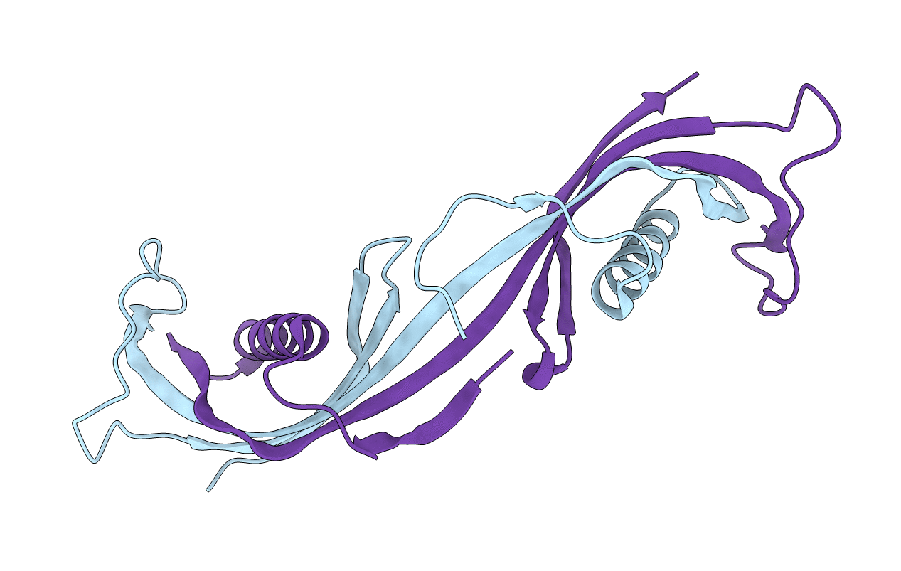
PDB ID:
1TIJ
Keywords:
Title:
3D Domain-swapped human cystatin C with amyloid-like intermolecular beta-sheets
Biological Source:
Source Organism(s):
Homo sapiens (Taxon ID: 9606)
Expression System(s):
Method Details:
Experimental Method:
Resolution:
3.03 Å
R-Value Free:
0.26
R-Value Work:
0.21
R-Value Observed:
0.21
Space Group:
P 41 21 2
