Deposition Date
2004-05-27
Release Date
2004-09-14
Last Version Date
2024-02-14
Entry Detail
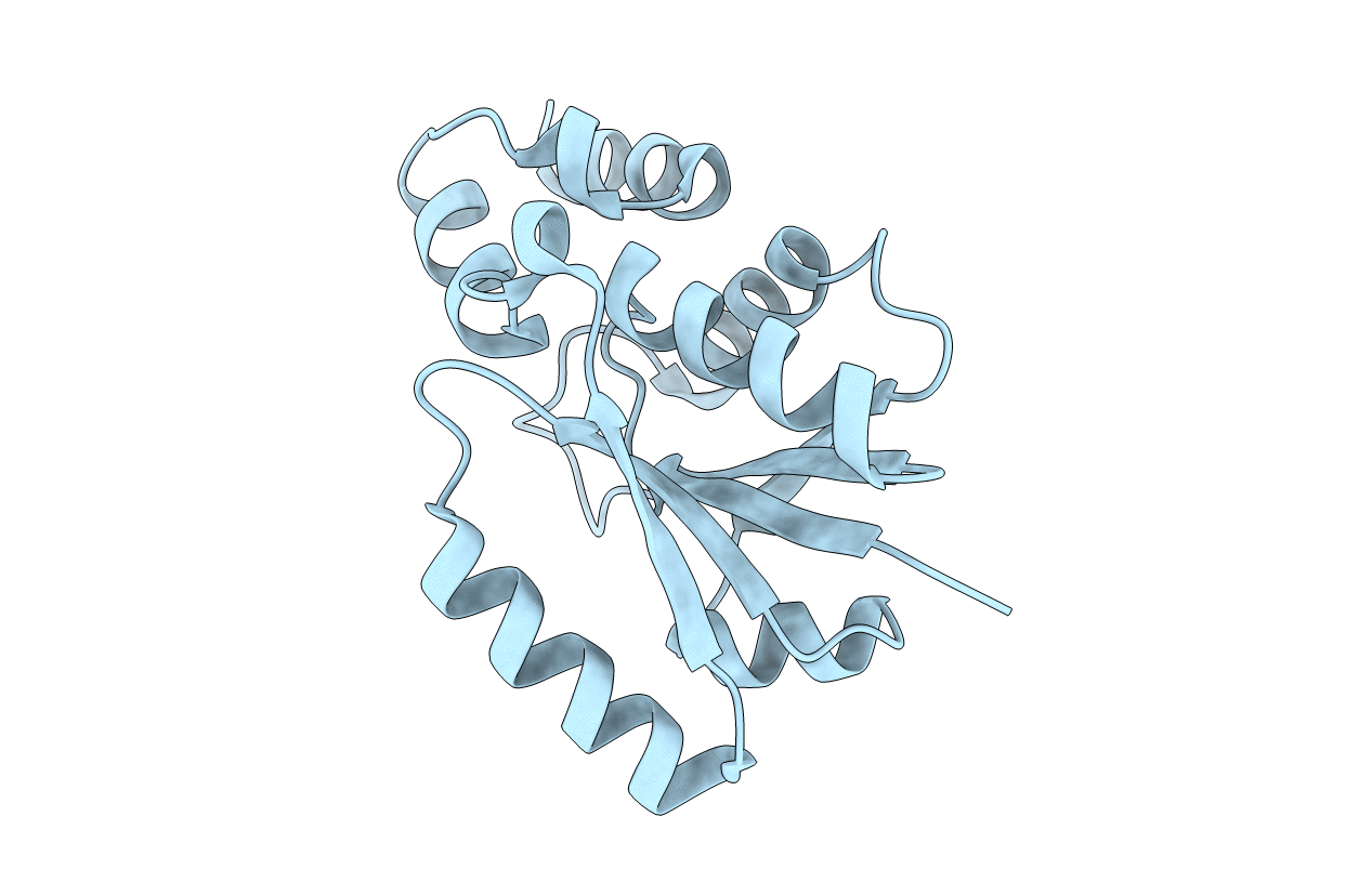
PDB ID:
1TFU
Keywords:
Title:
phosphopantetheine adenylyltransferase from Mycobacterium tuberculosis
Biological Source:
Source Organism(s):
Mycobacterium tuberculosis (Taxon ID: 1773)
Expression System(s):
Method Details:
Experimental Method:
Resolution:
1.99 Å
R-Value Free:
0.25
R-Value Work:
0.22
R-Value Observed:
0.22
Space Group:
H 3 2