Deposition Date
2004-05-26
Release Date
2004-08-24
Last Version Date
2024-02-14
Entry Detail
PDB ID:
1TF7
Keywords:
Title:
Crystal Structure of Circadian Clock Protein KaiC
Biological Source:
Source Organism(s):
Synechococcus sp. (Taxon ID: 1131)
Expression System(s):
Method Details:
Experimental Method:
Resolution:
2.80 Å
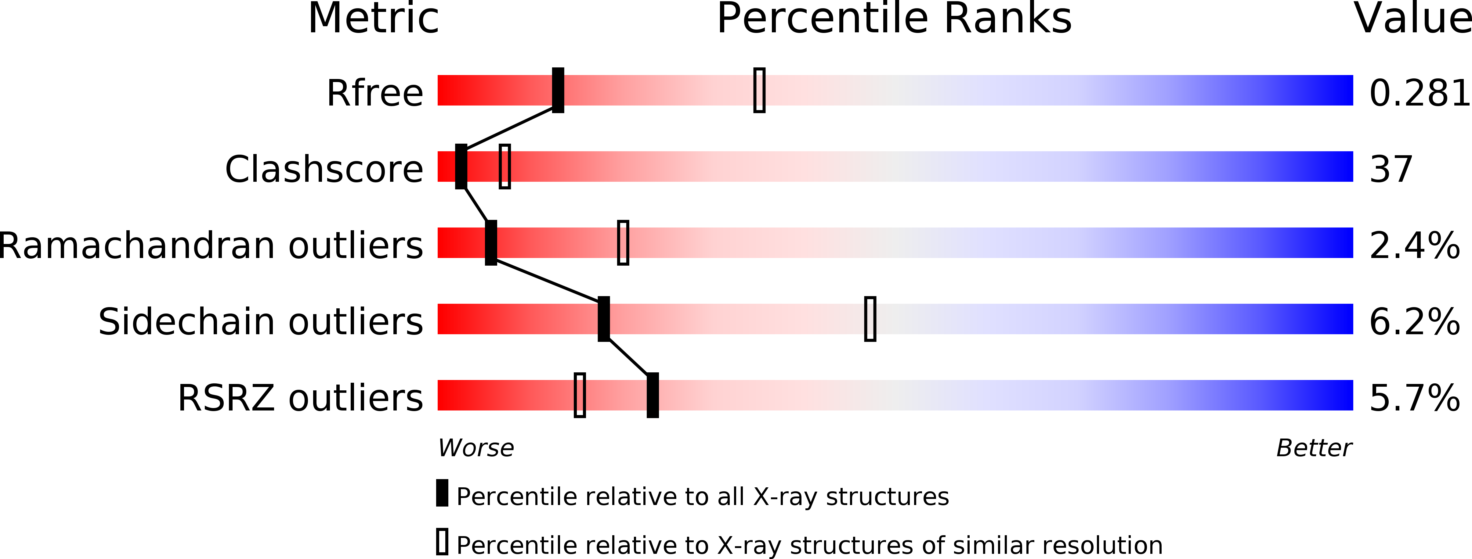
R-Value Free:
0.28
R-Value Work:
0.24
R-Value Observed:
0.24
Space Group:
P 21 21 21