Deposition Date
1994-04-08
Release Date
1995-02-07
Last Version Date
2024-11-20
Entry Detail
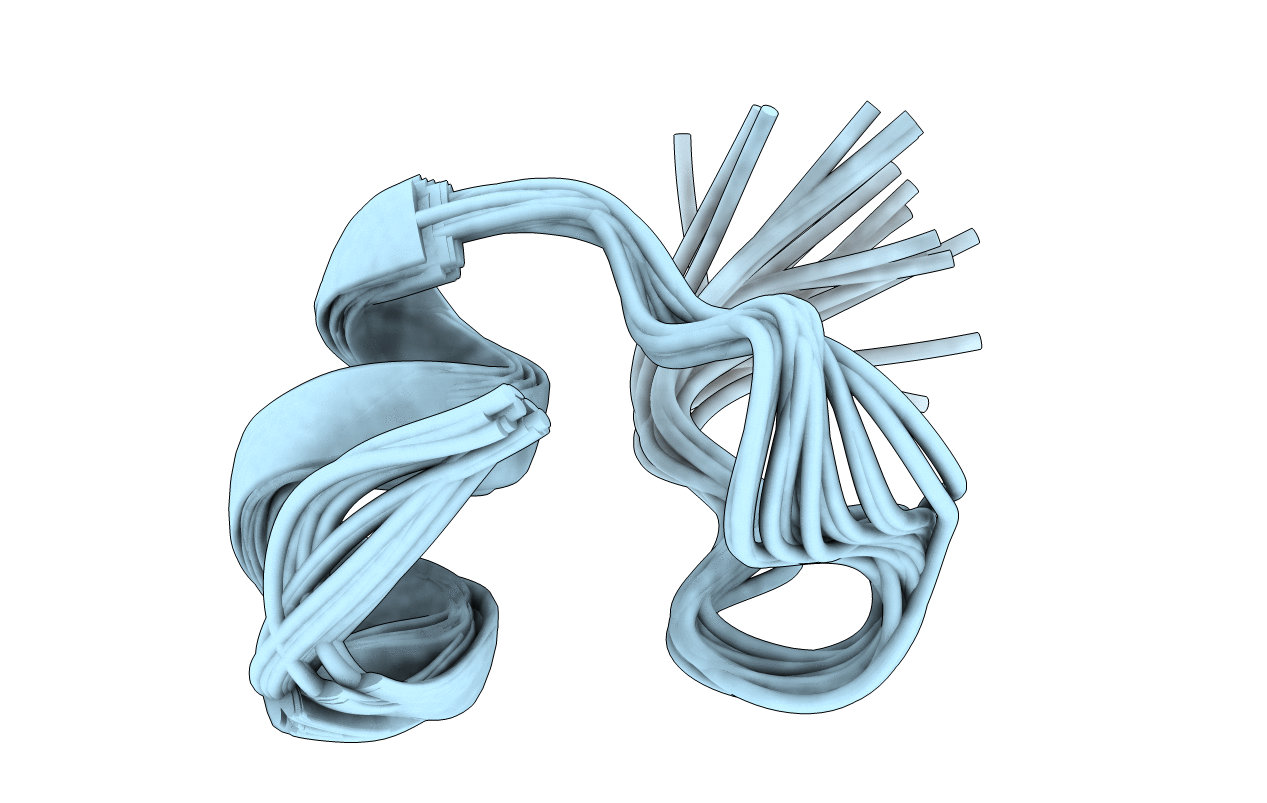
PDB ID:
1TER
Keywords:
Title:
SOLUTION STRUCTURE OF TERTIAPIN DETERMINED USING NUCLEAR MAGNETIC RESONANCE AND DISTANCE GEOMETRY
Biological Source:
Source Organism(s):
Apis mellifera (Taxon ID: 7460)
Method Details:
Experimental Method:
Conformers Submitted:
21