
Deposition Date
2004-05-25
Release Date
2004-08-03
Last Version Date
2024-04-03
Entry Detail
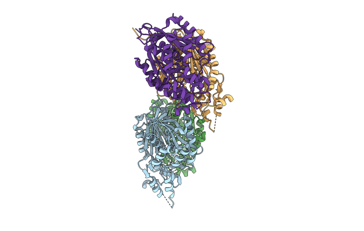
PDB ID:
1TEE
Keywords:
Title:
Crystal structure of C205F mutant of PKS18 from Mycobacterium tuberculosis
Biological Source:
Source Organism(s):
Mycobacterium tuberculosis (Taxon ID: 83332)
Expression System(s):
Method Details:
Experimental Method:
Resolution:
2.90 Å
R-Value Free:
0.25
R-Value Work:
0.22
R-Value Observed:
0.22
Space Group:
P 1
