
Deposition Date
2004-05-24
Release Date
2004-09-21
Last Version Date
2023-08-23
Entry Detail
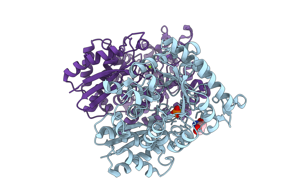
PDB ID:
1TE6
Keywords:
Title:
Crystal Structure of Human Neuron Specific Enolase at 1.8 angstrom
Biological Source:
Source Organism(s):
Homo sapiens (Taxon ID: 9606)
Expression System(s):
Method Details:
Experimental Method:
Resolution:
1.80 Å
R-Value Free:
0.23
R-Value Work:
0.20
R-Value Observed:
0.20
Space Group:
P 21 21 2
