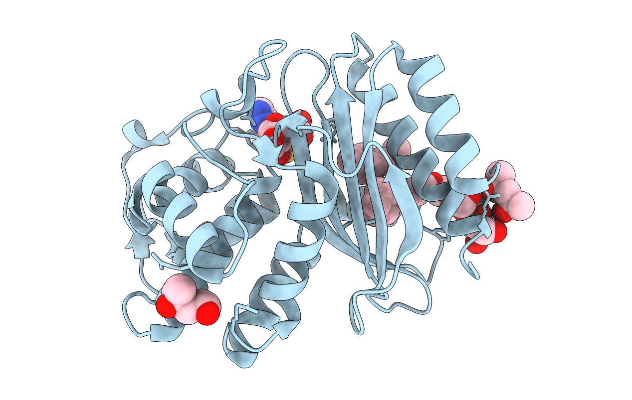
1TDG image
Deposition Date 2004-05-21
Release Date 2004-11-23
Last Version Date 2024-11-20
Entry Detail
PDB ID:
1TDG
Keywords:
Title:
Complex of S130G SHV-1 beta-lactamase with tazobactam
Biological Source:
Source Organism:
Method Details:
Experimental Method:
Resolution:
1.80 Å
R-Value Free:
0.17
R-Value Work:
0.14
R-Value Observed:
0.14
Space Group:
P 21 21 21
Macromolecular Entities
Polymer Type:polypeptide(L)
Molecule:Beta-lactamase SHV-1
Mutations:S130G
Chain IDs:A
Chain Length:265
Number of Molecules:1
Biological Source:Klebsiella pneumoniae
Primary Citation
Inhibitor-resistant class A beta-lactamases: consequences of the Ser130-to-Gly mutation seen in Apo and tazobactam structures of the SHV-1 variant
Biochemistry 43 14111 14117 (2004)
PMID: 15518561 DOI: 10.1021/bi0487903

Abstact

A bacterial response to the clinical use of class A beta-lactamase inhibitors such as tazobactam and clavulanic acid is the expression of variant beta-lactamases with weaker binding affinities for these mechanism-based inhibitors. Some of these inhibitor-resistant variants contain a glycine mutation at Ser130, a conserved active site residue known to be adventitiously involved in the inhibition mechanism. The crystallographic structure of a complex of tazobactam with the Ser130Gly variant of the class A SHV-1 beta-lactamase has been determined to 1.8 A resolution. Two reaction intermediates are observed. The primary intermediate is an acyclic species bound to the reactive Ser70. It is poorly primed for catalytic hydrolysis because its ester carbonyl group is completely displaced from the enzyme's oxyanion hole. A smaller fraction of the enzyme contains a Ser70-bound aldehyde resulting from hydrolytic loss of the triazoyl-sulfinyl amino acid moiety from the primary species. This first structure of a class A beta-lactamase lacking Ser130, the side chain of which functions in beta-lactam binding and possibly in catalysis, gives crystallographic evidence that the acylation step of beta-lactam turnover can occur without Ser130. Unexpectedly, the crystal structure of the uncomplexed Ser130Gly enzyme, also determined to 1.8 A resolution, shows that a critical Glu166-activated water molecule is missing from the catalytic site. Comparison of this uncomplexed variant with the wild-type structure reveals that Ser130 is required for orienting the side chain of Ser70 and ensuring the hydrogen bonding of Ser70 to both Lys73 and the catalytic water molecule.

Legend

Protein

Chemical

Disease

Primary Citation of related structures