1TB3 image
Entry Detail
PDB ID:
1TB3
Keywords:
Title:
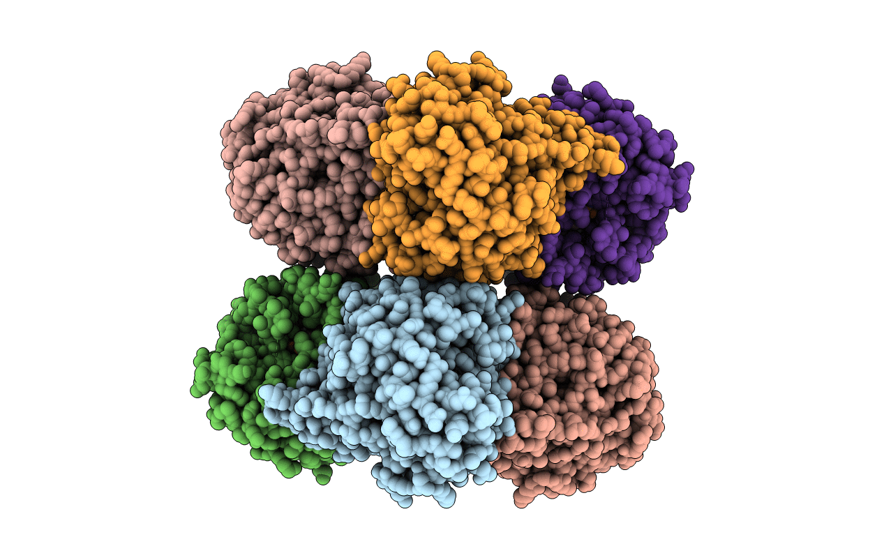
Crystal Structure Analysis of Recombinant Rat Kidney Long-chain Hydroxy Acid Oxidase
Biological Source:
Source Organism:
Host Organism:
PDB Version:
Deposition Date:
2004-05-19
Release Date:
2005-02-01
Method Details:
Experimental Method:
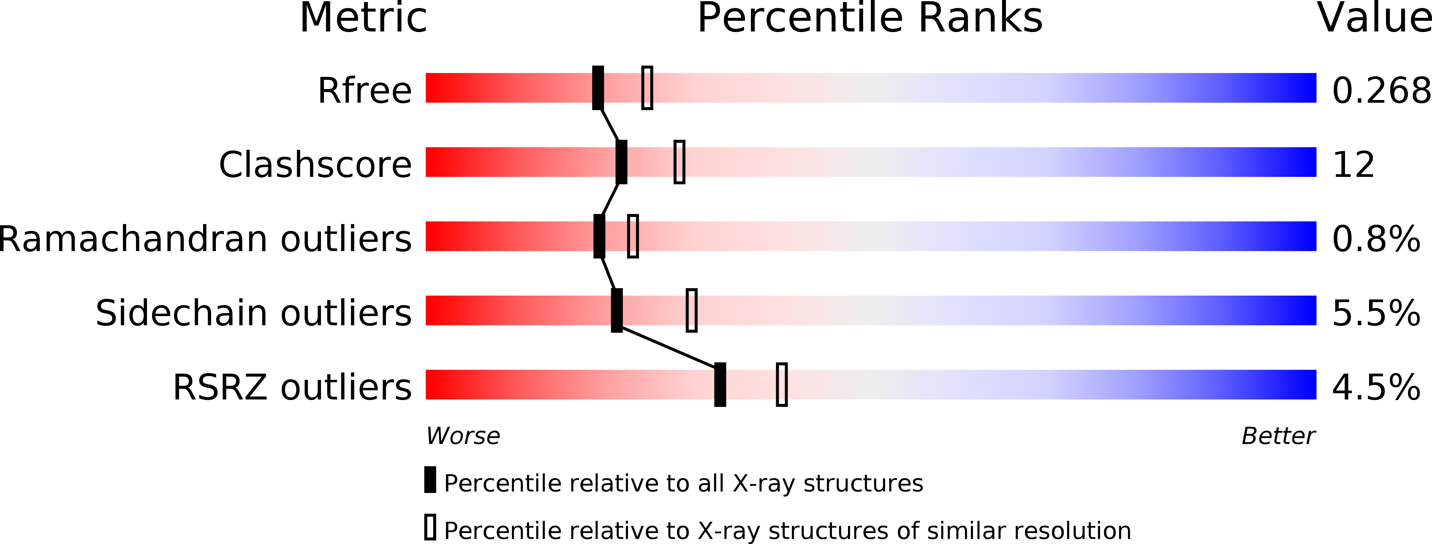
Resolution:
2.30 Å
R-Value Free:
0.26
R-Value Work:
0.23
Space Group:
P 21 21 21
Macromolecular Entities
Polymer Type:polypeptide(L)
Description:Hydroxyacid oxidase 3
Chain IDs:A, B, C, D, E, F, G, H
Chain Length:352
Number of Molecules:8
Biological Source:Rattus norvegicus
Primary Citation
Crystal Structure Analysis of Recombinant Rat Kidney Long Chain Hydroxy Acid Oxidase.
Biochemistry 44 1521 1531 (2005)
PMID: 15683236 DOI: 10.1021/bi048616e

Abstact

Long chain hydroxy acid oxidase (LCHAO) is a member of an FMN-dependent enzyme family that oxidizes L-2-hydroxy acids to ketoacids. LCHAO is a peroxisomal enzyme, and the identity of its physiological substrate is unclear. Mandelate is the most efficient substrate known and is commonly used in the test tube. LCHAO differs from most family members in that one of the otherwise invariant active site residues is a phenylalanine (Phe23) instead of a tyrosine. We now report the crystal structure of LCHAO. It shows the same beta8alpha8 TIM barrel structure as other structurally characterized family members, e.g., spinach glycolate oxidase (GOX) and the electron transferases yeast flavocytochrome b2 (FCB2) and Pseudomonas putida mandelate dehydrogenase (MDH). Loop 4, which is mobile in other family members, is visible in part. An acetate ion is present in the active site. The flavin interacts with the protein in the same way as in the electron transferases, and not as in GOX, an unexpected observation. An interpretation is proposed to explain this difference between GOX on one hand and FCB2 and LCHAO on the other hand, which had been proposed to arise from the differences between family members in their reactivity with oxygen. A comparison of models of the substrate bound to various published structures suggests that the very different reactivity with mandelate of LCHAO, GOX, FCB2, and MDH cannot be rationalized by a hydride transfer mechanism.

Legend

Protein

Chemical

Disease

Primary Citation of related structures