Deposition Date
1993-10-04
Release Date
1994-01-31
Last Version Date
2024-02-14
Entry Detail
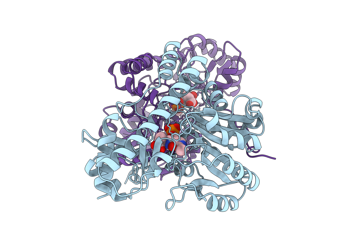
PDB ID:
1TAS
Keywords:
Title:
CRYSTALLINE MITOCHONDRIAL ASPARTATE AMINOTRANSFERASE EXISTS IN ONLY TWO CONFORMATIONS
Biological Source:
Source Organism(s):
Gallus gallus (Taxon ID: 9031)
Expression System(s):
Method Details:
Experimental Method:
Resolution:
2.80 Å
R-Value Observed:
0.16
Space Group:
P 1 21 1