1TAP image
Deposition Date 1994-08-16
Release Date 1994-11-30
Last Version Date 2024-11-20
Entry Detail
PDB ID:
1TAP
Title:
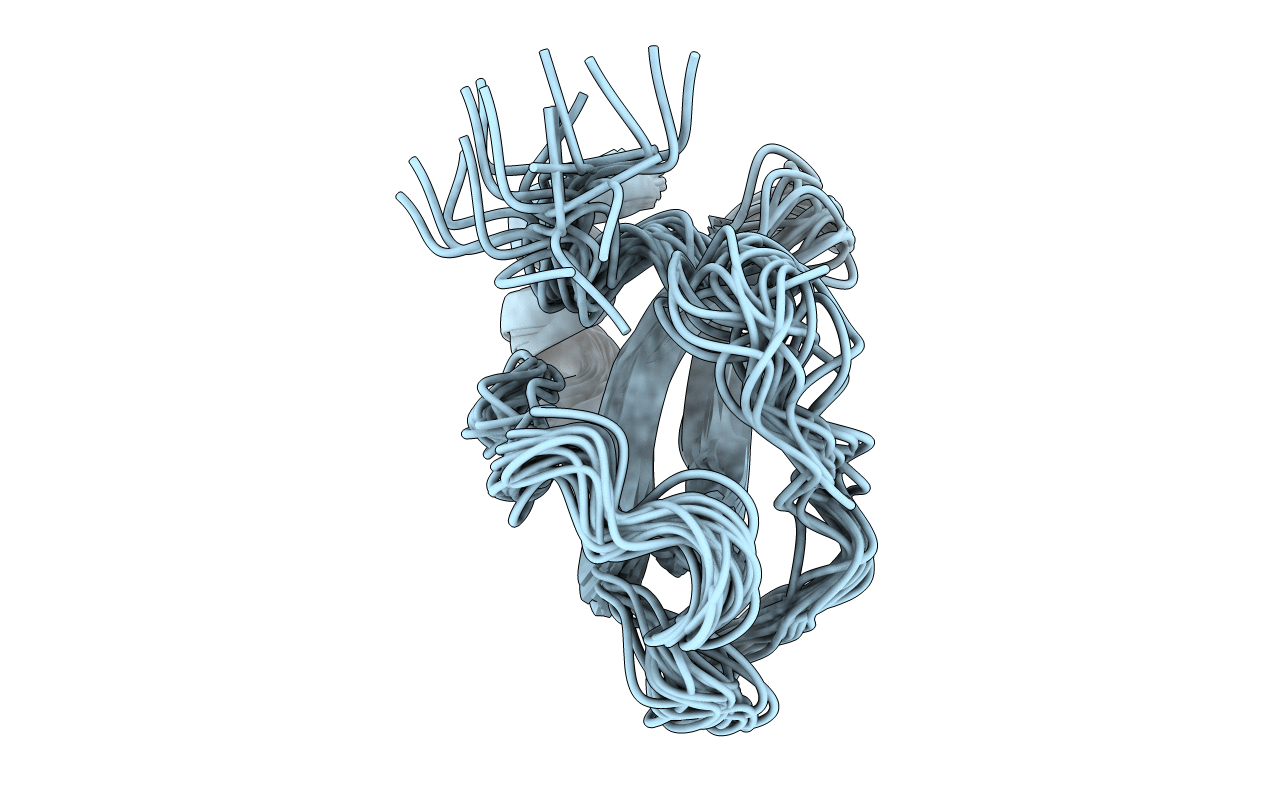
NMR SOLUTION STRUCTURE OF RECOMBINANT TICK ANTICOAGULANT PROTEIN (RTAP), A FACTOR XA INHIBITOR FROM THE TICK ORNITHODOROS MOUBATA
Biological Source:
Source Organism:
Method Details:
Experimental Method:
Conformers Submitted:
20
Macromolecular Entities
Polymer Type:polypeptide(L)
Molecule:FACTOR XA INHIBITOR
Chain IDs:A
Chain Length:60
Number of Molecules:1
Biological Source:Ornithodoros moubata
Ligand Molecules
Primary Citation
NMR solution structure of the recombinant tick anticoagulant protein (rTAP), a factor Xa inhibitor from the tick Ornithodoros moubata.
FEBS Lett. 352 251 257 (1994)
PMID: 7925983 DOI: 10.1016/0014-5793(94)00941-4

Abstact

The solution structure of the recombinant tick anticoagulant protein (rTAP) was determined by 1H nuclear magnetic resonance (NMR) spectroscopy in aqueous solution at pH 3.6 and 36 degrees C. rTAP is a 60-residue protein functioning as a highly specific inhibitor of the coagulation protease factor Xa, which was originally isolated from the tick Ornithodoros moubata. Its regular secondary structure consists of a two-stranded antiparallel beta-sheet with residues 22-28 and 32-38, and an alpha-helix with residues 51-60. The relative orientation of these regular secondary structure elements has nearly identical counterparts in the bovine pancreatic trypsin inhibitor (BPTI). In contrast, the loop between the beta-sheet and the C-terminal alpha-helix as well as the N-terminal 20-residue segment preceding the beta-sheet adopt different three-dimensional folds in the two proteins. These observations are discussed with regard to the implication of different mechanisms of protease inhibition by rTAP and by Kunitz-type protein proteinase inhibitors.

Legend

Protein

Chemical

Disease

Primary Citation of related structures