
Deposition Date
2004-05-13
Release Date
2004-08-17
Last Version Date
2024-10-16
Entry Detail
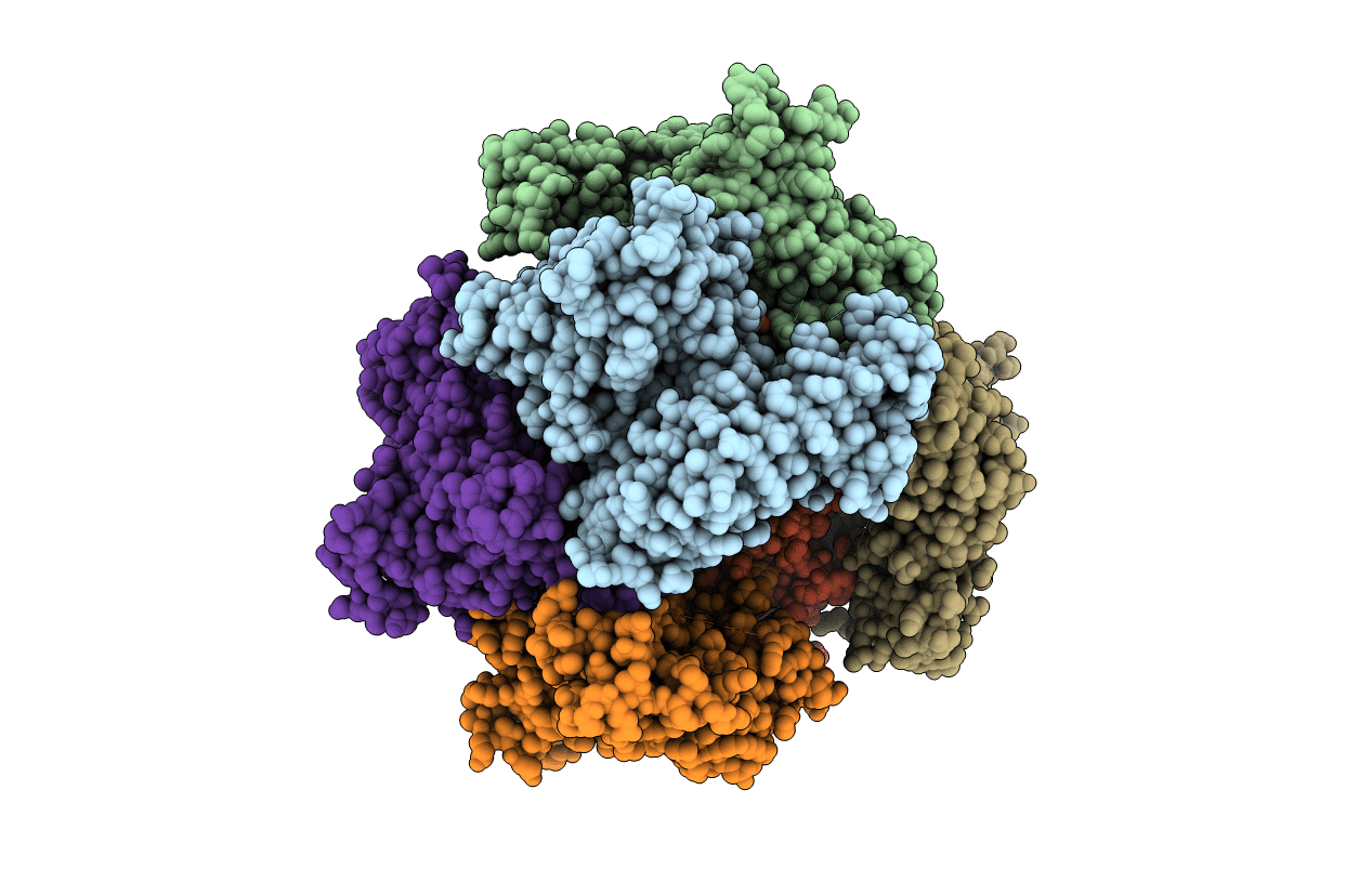
PDB ID:
1T8S
Keywords:
Title:
Crystal Structure of E.coli AMP Nucleosidase complexed with formicin 5'-monophosphate
Biological Source:
Source Organism(s):
Escherichia coli (Taxon ID: 562)
Expression System(s):
Method Details:
Experimental Method:
Resolution:
2.60 Å
R-Value Free:
0.25
R-Value Work:
0.22
R-Value Observed:
0.22
Space Group:
P 42 21 2
