
Deposition Date
2004-05-13
Release Date
2004-08-10
Last Version Date
2023-10-25
Entry Detail
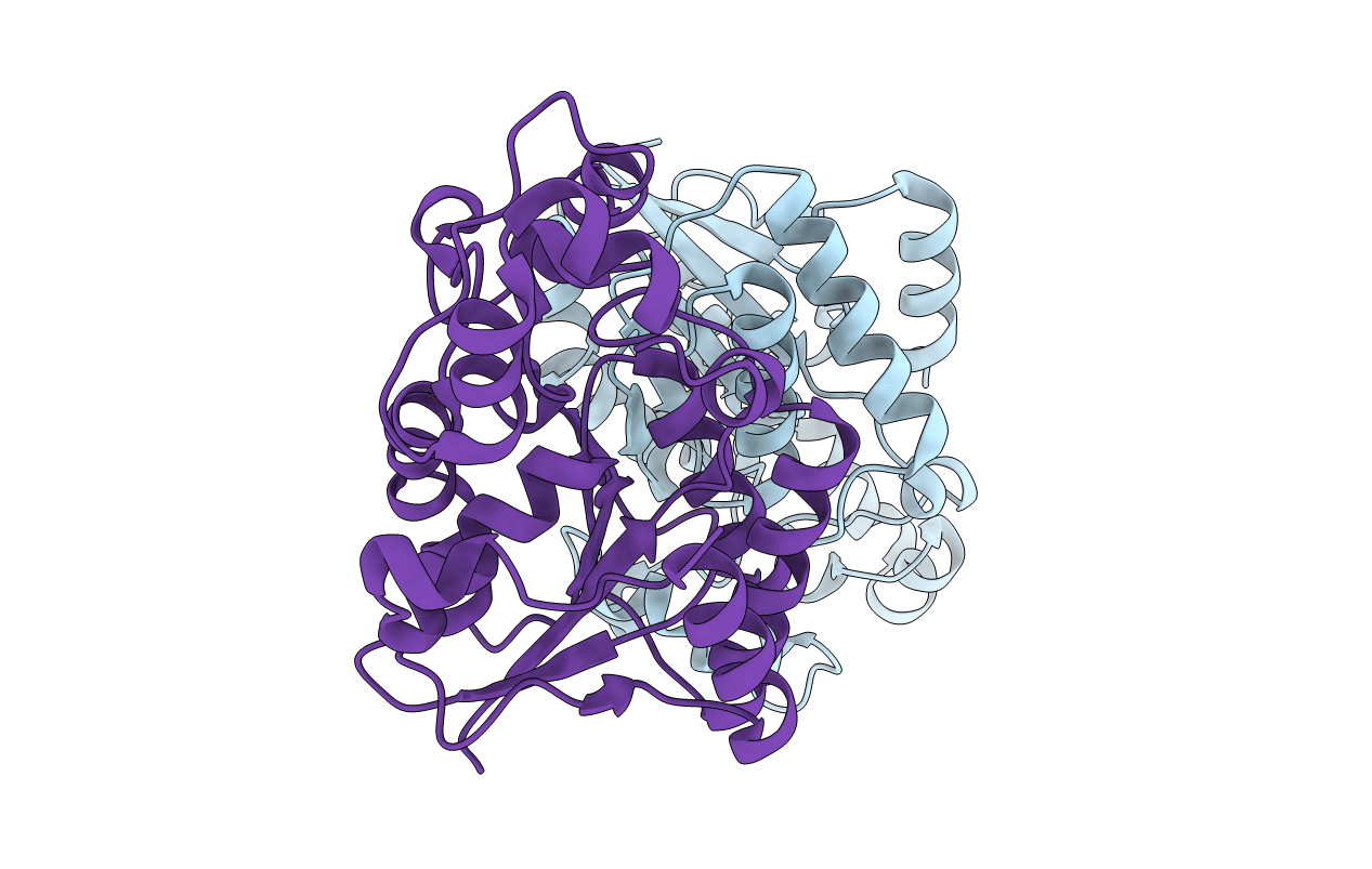
PDB ID:
1T8P
Keywords:
Title:
Crystal structure of Human erythrocyte 2,3-bisphosphoglycerate mutase
Biological Source:
Source Organism(s):
Homo sapiens (Taxon ID: 9606)
Expression System(s):
Method Details:
Experimental Method:
Resolution:
2.50 Å
R-Value Free:
0.26
R-Value Work:
0.2
R-Value Observed:
0.2
Space Group:
P 1 21 1
