1T8B image
Deposition Date 2004-05-11
Release Date 2004-12-07
Last Version Date 2023-08-23
Entry Detail
PDB ID:
1T8B
Title:
Crystal structure of refolded PHOU-like protein (gi 2983430) from Aquifex aeolicus
Biological Source:
Source Organism:
Host Organism:
Method Details:
Experimental Method:
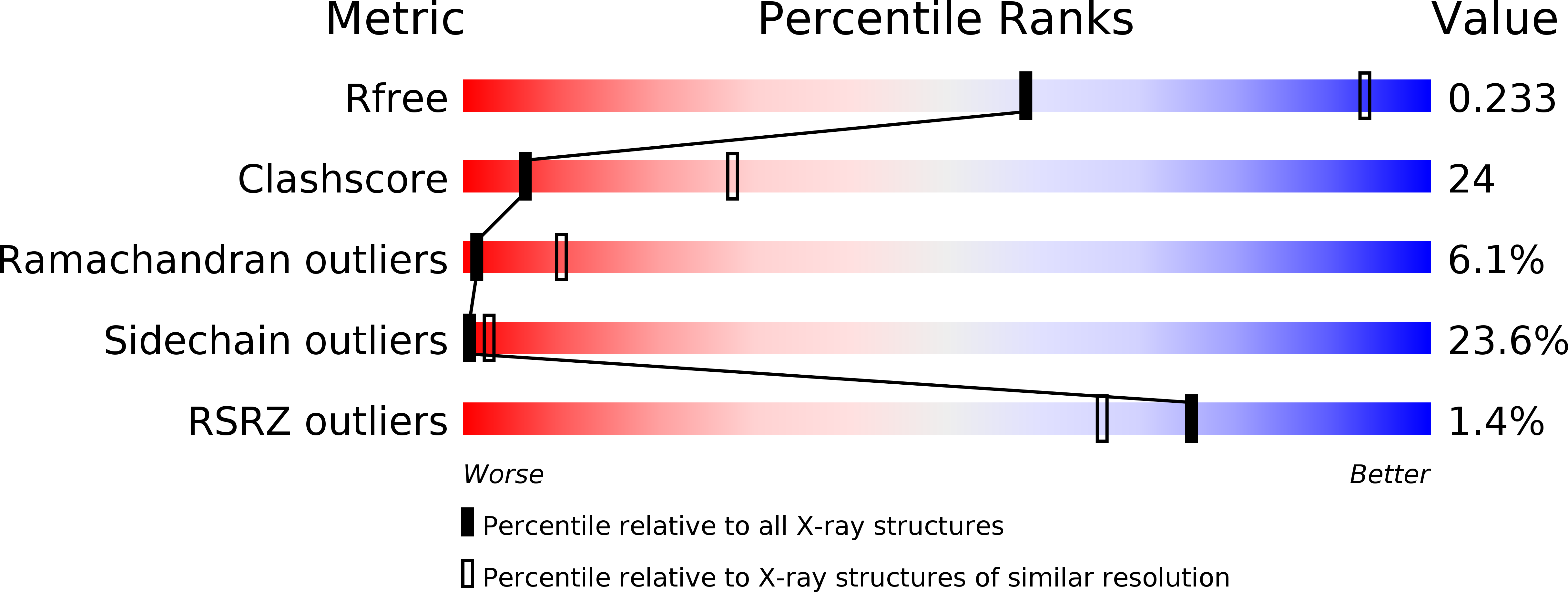
Resolution:
3.23 Å
R-Value Free:
0.24
R-Value Work:
0.21
R-Value Observed:
0.21
Space Group:
P 32
Macromolecular Entities
Polymer Type:polypeptide(L)
Molecule:Phosphate transport system protein phoU homolog
Gene (Uniprot):phoU
Chain IDs:A, B
Chain Length:227
Number of Molecules:2
Biological Source:Aquifex aeolicus
Ligand Molecules
Primary Citation
Crystal structure of the "PhoU-like" phosphate uptake regulator from Aquifex aeolicus.
J.Bacteriol. 187 4238 4244 (2005)
PMID: 15937186 DOI: 10.1128/JB.187.12.4238-4244.2005

Abstact

The phoU gene of Aquifex aeolicus encodes a protein called PHOU_AQUAE with sequence similarity to the PhoU protein of Escherichia coli. Despite the fact that there is a large number of family members (more than 300) attributed to almost all known bacteria and despite PHOU_AQUAE's association with the regulation of genes for phosphate metabolism, the nature of its regulatory function is not well understood. Nearly one-half of these PhoU-like proteins, including both PHOU_AQUAE and the one from E. coli, form a subfamily with an apparent dimer structure of two PhoU domains on the basis of their amino acid sequence. The crystal structure of PHOU_AQUAE (a 221-amino-acid protein) reveals two similar coiled-coil PhoU domains, each forming a three-helix bundle. The structures of PHOU_AQUAE proteins from both a soluble fraction and refolded inclusion bodies (at resolutions of 2.8 and 3.2A, respectively) showed no significant differences. The folds of the PhoU domain and Bag domains (for a class of cofactors of the eukaryotic chaperone Hsp70 family) are similar. Accordingly, we propose that gene regulation by PhoU may occur by association of PHOU_AQUAE with the ATPase domain of the histidine kinase PhoR, promoting release of its substrate PhoB. Other proteins that share the PhoU domain fold include the coiled-coil domains of the STAT protein, the ribosome-recycling factor, and structural proteins like spectrin.

Legend

Protein

Chemical

Disease

Primary Citation of related structures