Deposition Date
2004-05-10
Release Date
2004-05-18
Last Version Date
2024-11-06
Entry Detail
PDB ID:
1T7S
Keywords:
Title:
Structural Genomics of Caenorhabditis elegans: Structure of BAG-1 protein
Biological Source:
Source Organism(s):
Caenorhabditis elegans (Taxon ID: 6239)
Expression System(s):
Method Details:
Experimental Method:
Resolution:
2.80 Å
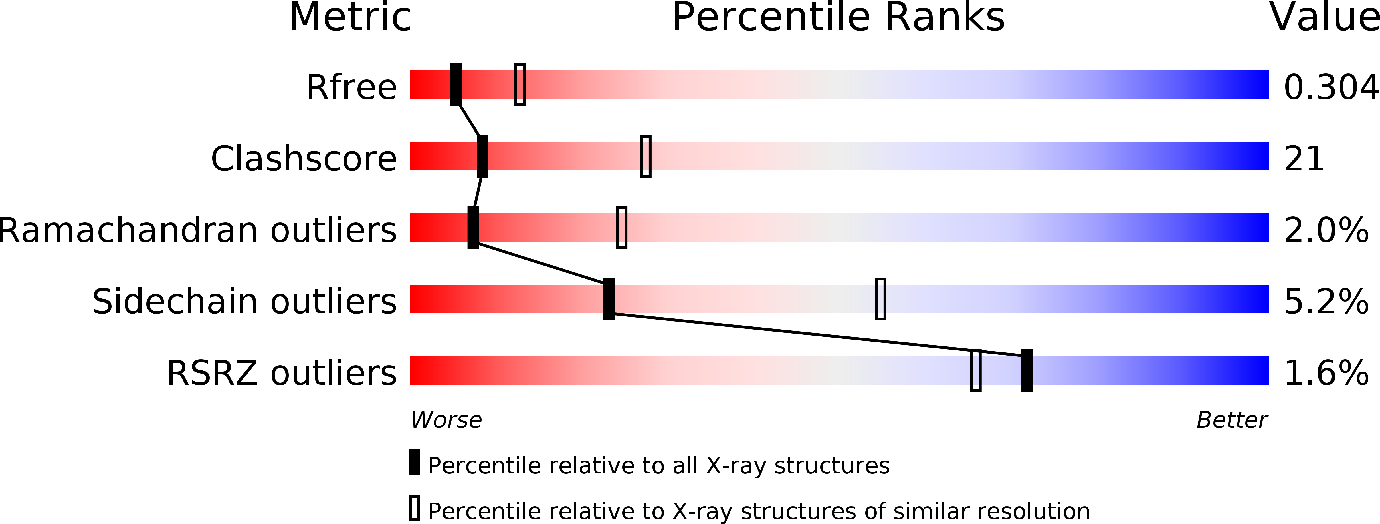
R-Value Free:
0.29
R-Value Work:
0.22
R-Value Observed:
0.22
Space Group:
I 2 2 2