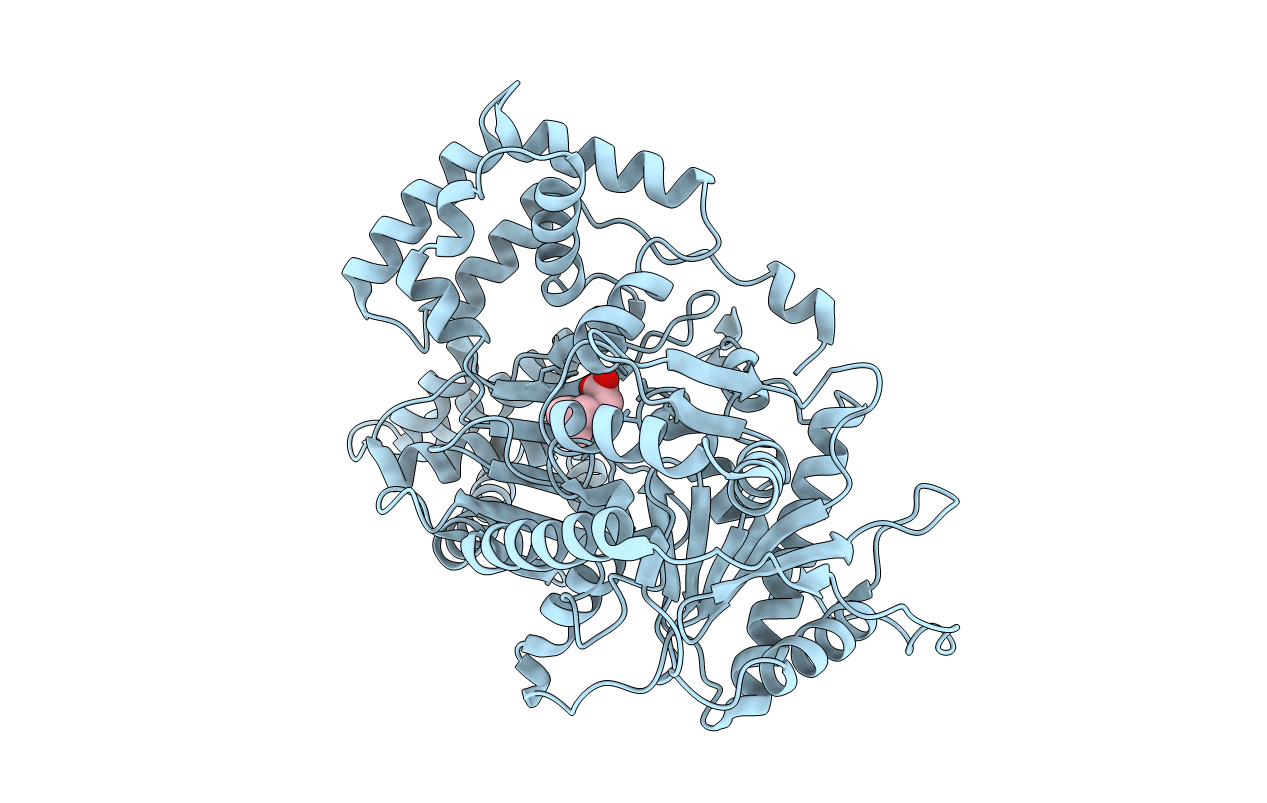
1T7O image
Entry Detail
PDB ID:
1T7O
Keywords:
Title:
Crystal structure of the M564G mutant of murine carnitine acetyltransferase in complex with carnitine
Biological Source:
Source Organism:
Host Organism:
PDB Version:
Deposition Date:
2004-05-10
Release Date:
2004-06-22
Method Details:
Experimental Method:
Resolution:
2.30 Å
R-Value Free:
0.26
R-Value Work:
0.20
R-Value Observed:
0.20
Space Group:
P 43
Macromolecular Entities
Polymer Type:polypeptide(L)
Description:Carnitine acetyltransferase
Mutations:M564G
Chain IDs:A
Chain Length:618
Number of Molecules:1
Biological Source:Mus musculus
Ligand Molecules
Primary Citation
Structural and biochemical studies of the substrate selectivity of carnitine acetyltransferase
J.Biol.Chem. 279 31584 31589 (2004)
PMID: 15155726 DOI: 10.1074/jbc.M403484200

Abstact

Carnitine acyltransferases catalyze the exchange of acyl groups between coenzyme A (CoA) and carnitine. They have important roles in many cellular processes, especially the oxidation of long-chain fatty acids, and are attractive targets for drug discovery against diabetes and obesity. These enzymes are classified based on their substrate selectivity for short-chain, medium-chain, or long-chain fatty acids. Structural information on carnitine acetyltransferase suggests that residues Met-564 and Phe-565 may be important determinants of substrate selectivity with the side chain of Met-564 located in the putative binding pocket for acyl groups. Both residues are replaced by glycine in carnitine palmitoyltransferases. To assess the functional relevance of this structural observation, we have replaced these two residues with small amino acids by mutagenesis, characterized the substrate preference of the mutants, and determined the crystal structures of two of these mutants. Kinetic studies confirm that the M564G or M564A mutation is sufficient to increase the activity of the enzyme toward medium-chain substrates with hexanoyl-CoA being the preferred substrate for the M564G mutant. The crystal structures of the M564G mutant, both alone and in complex with carnitine, reveal a deep binding pocket that can accommodate the larger acyl group. We have determined the crystal structure of the F565A mutant in a ternary complex with both the carnitine and CoA substrates at a 1.8-A resolution. The F565A mutation has minor effects on the structure or the substrate preference of the enzyme.

Legend

Protein

Chemical

Disease

Primary Citation of related structures