1T7H image
Deposition Date 2004-05-10
Release Date 2004-12-21
Last Version Date 2024-11-06
Entry Detail
PDB ID:
1T7H
Title:
X-ray structure of [Lys(-2)-Arg(-1)-des(17-21)]-endothelin-1 peptide
Biological Source:
Source Organism:
Homo sapiens (Taxon ID: 9606)
Method Details:
Experimental Method:
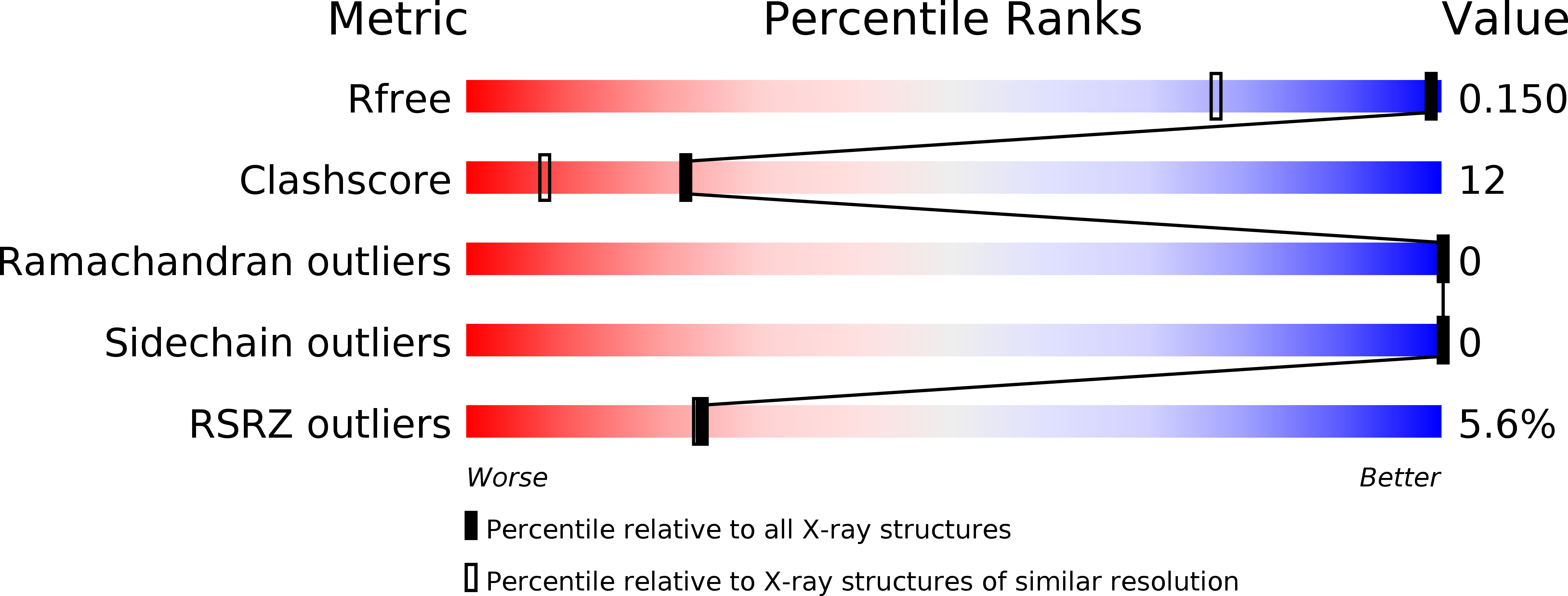
Resolution:
1.13 Å
R-Value Free:
0.13
R-Value Work:
0.12
R-Value Observed:
0.12
Space Group:
P 21 21 2
Macromolecular Entities
Polymer Type:polypeptide(L)
Molecule:Endothelin-1
Gene (Uniprot):EDN1
Chain IDs:A, B
Chain Length:18
Number of Molecules:2
Biological Source:Homo sapiens
Primary Citation
High-resolution X-ray structure of the unexpectedly stable dimer of the [Lys(-2)-Arg(-1)-des(17-21)]endothelin-1 peptide
Biochemistry 43 15154 15168 (2004)
PMID: 15568807 DOI: 10.1021/bi049098a

Abstact

Previous structural studies on the [Lys((-2))-Arg((-1))]endothelin-1 peptide (KR-ET-1), 540-fold less potent than ET-1, strongly suggested the presence of an intramolecular Arg(-1)-Asp(8) (R(-1)-D(8)) salt bridge that was also observed in the shorter [Lys((-2))-Arg((-1))-des(17-21)]endothelin-1 derivative (KR-CSH-ET). In addition, for these two analogues, we have shown that the Lys-Arg dipeptide, which belongs to the prosequence, significantly improves the formation of the native disulfide bonds (>or=96% instead of approximately 70% for ET-1). In contrast to what was inferred from NMR data, molecular dynamics simulations suggested that such an intramolecular salt bridge would be unstable. The KR-CSH-ET peptide has now been crystallized at pH 5.0 and its high-resolution structure determined ab initio at 1.13 A using direct methods. Unexpectedly, KR-CSH-ET was shown to be a head-to-tail symmetric dimer, and the overall interface involves two intermolecular R(-1)-D(8) salt bridges, a two-stranded antiparallel beta-sheet, and hydrophobic contacts. Molecular dynamics simulations carried out on this dimer clearly showed that the two intermolecular salt bridges were in this case very stable. Sedimentation equilibrium experiments unambiguously confirmed that KR-ET-1 and KR-CSH-ET also exist as dimers in solution at pH 5.0. On the basis of the new dimeric structure, previous NMR data were reinterpreted. Structure calculations were performed using 484 intramolecular and 38 intermolecular NMR-derived constraints. The solution and the X-ray structures of the dimer are very similar (mean rmsd of 0.85 A). Since the KR dipeptide at the N-terminus of KR-CSH-ET is present in the prosequence, it can be hypothesized that similar intermolecular salt bridges could be involved in the in vivo formation of the native disulfide bonds of ET-1. Therefore, it appears to be likely that the prosequence does assist the ET-1 folding in a chaperone-like manner before successive cleavages that yield the bioactive ET-1 hormone.

Legend

Protein

Chemical

Disease

Primary Citation of related structures