1T6V image
Entry Detail
PDB ID:
1T6V
Title:
Crystal structure analysis of the nurse shark new antigen receptor (NAR) variable domain in complex with lysozyme
Biological Source:
Host Organism:
PDB Version:
Deposition Date:
2004-05-07
Release Date:
2004-08-24
Method Details:
Experimental Method:
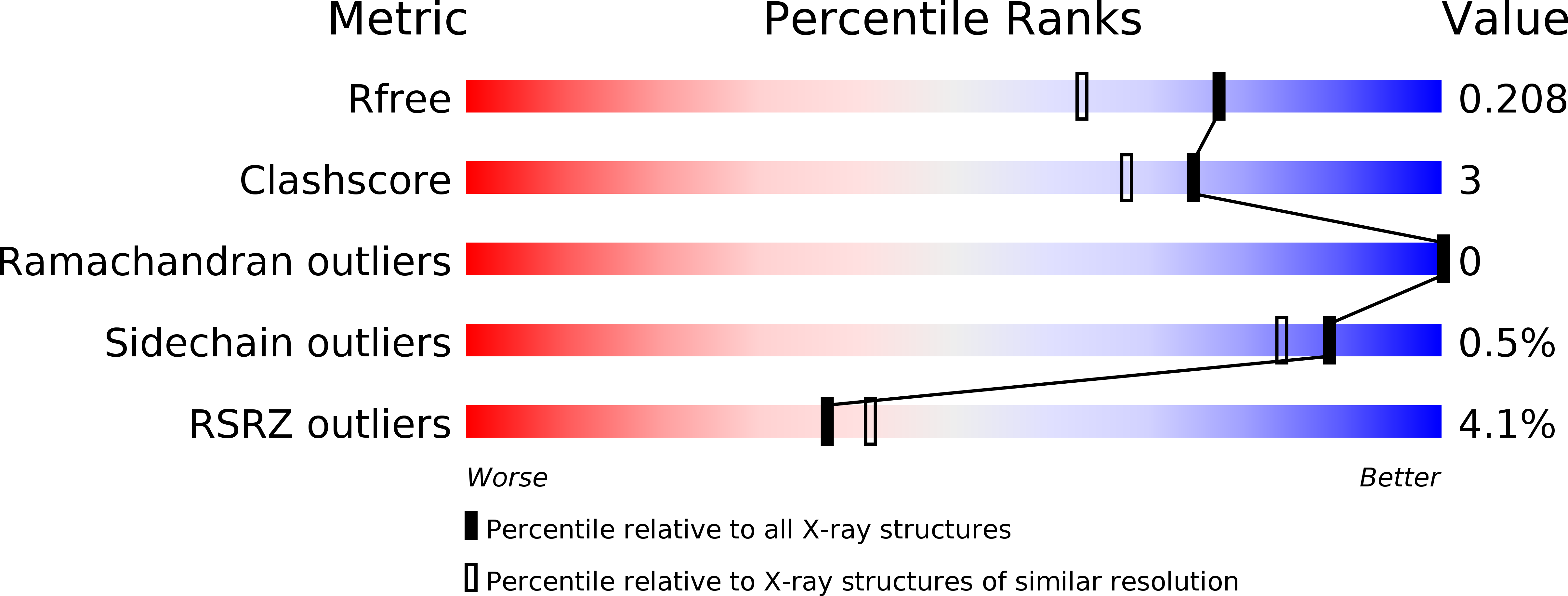
Resolution:
1.70 Å
R-Value Free:
0.22
R-Value Work:
0.19
R-Value Observed:
0.19
Space Group:
P 21 21 2
Macromolecular Entities
Polymer Type:polypeptide(L)
Description:Lysozyme C
Chain IDs:A (auth: L), C (auth: M)
Chain Length:129
Number of Molecules:2
Biological Source:Gallus gallus
Polymer Type:polypeptide(L)
Description:novel antigen receptor
Chain IDs:B (auth: N), D (auth: O)
Chain Length:113
Number of Molecules:2
Biological Source:Ginglymostoma cirratum
Ligand Molecules
Primary Citation
Crystal structure of a shark single-domain antibody V region in complex with lysozyme.
Science 305 1770 1773 (2004)
PMID: 15319492 DOI: 10.1126/science.1101148

Abstact

Cartilaginous fish are the phylogenetically oldest living organisms known to possess components of the vertebrate adaptive immune system. Key to their immune response are heavy-chain, homodimeric immunoglobulins called new antigen receptors (IgNARs), in which the variable (V) domains recognize antigens with only a single immunoglobulin domain, akin to camelid heavy-chain V domains. The 1.45 angstrom resolution crystal structure of the type I IgNAR V domain in complex with hen egg-white lysozyme (HEL) reveals a minimal antigen-binding domain that contains only two of the three conventional complementarity-determining regions but still binds HEL with nanomolar affinity by means of a binding interface comparable in size to conventional antibodies.

Legend

Protein

Chemical

Disease

Primary Citation of related structures