Deposition Date
2004-05-05
Release Date
2004-07-20
Last Version Date
2024-02-14
Entry Detail
PDB ID:
1T6B
Keywords:
Title:
Crystal structure of B. anthracis Protective Antigen complexed with human Anthrax toxin receptor
Biological Source:
Source Organism(s):
Bacillus anthracis (Taxon ID: 1392)
Homo sapiens (Taxon ID: 9606)
Homo sapiens (Taxon ID: 9606)
Expression System(s):
Method Details:
Experimental Method:
Resolution:
2.50 Å
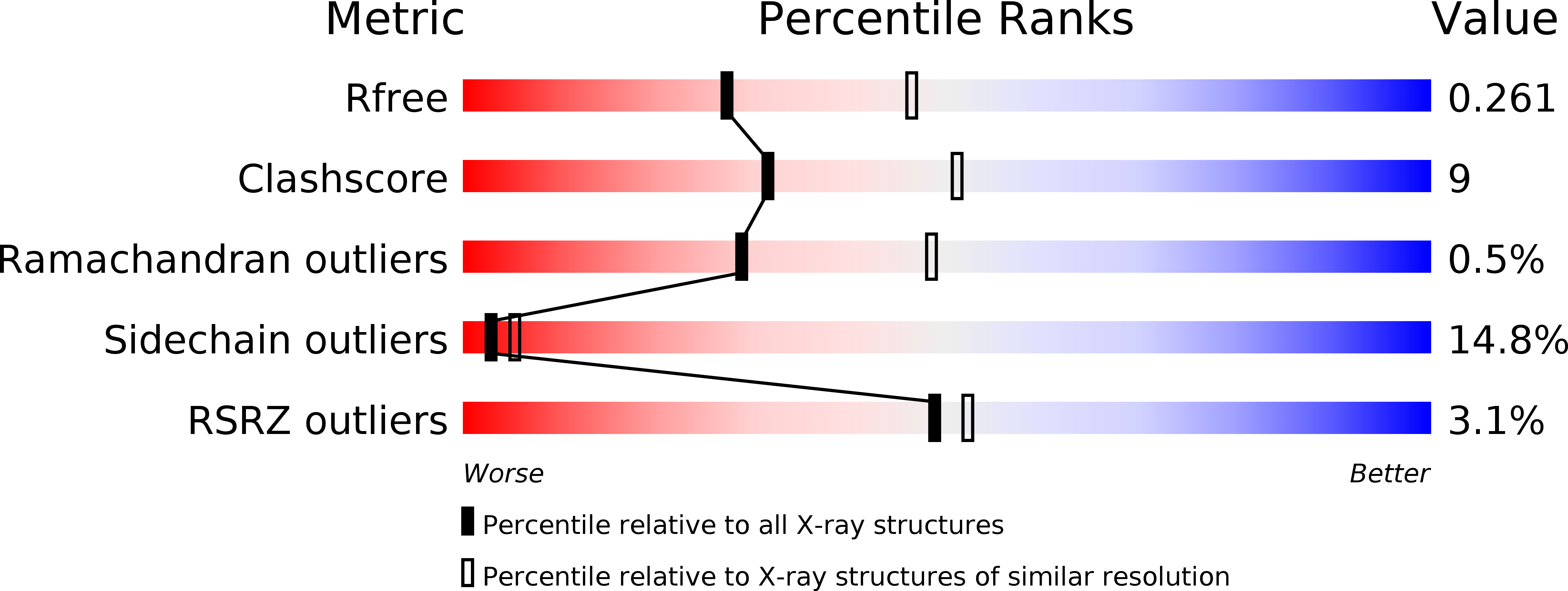
R-Value Free:
0.26
R-Value Work:
0.20
R-Value Observed:
0.21
Space Group:
P 21 21 21