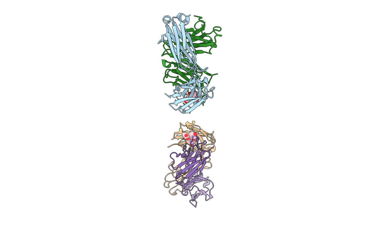
1T66 image
Deposition Date 2004-05-05
Release Date 2004-05-18
Last Version Date 2024-10-30
Entry Detail
PDB ID:
1T66
Keywords:
Title:
The structure of FAB with intermediate affinity for fluorescein.
Biological Source:
Source Organism:
Mus musculus (Taxon ID: 10090)
Method Details:
Experimental Method:
Resolution:
2.30 Å
R-Value Free:
0.25
R-Value Work:
0.20
R-Value Observed:
0.20
Space Group:
P 21 21 21
Macromolecular Entities
Structures with similar UniProt ID
Protein Blast
Polymer Type:polypeptide(L)
Molecule:immunoglobulin heavy chain
Chain IDs:B (auth: H), D
Chain Length:220
Number of Molecules:2
Biological Source:Mus musculus
Structures with similar UniProt ID
Protein Blast
Polymer Type:polypeptide(L)
Molecule:immunoglobulin light chain
Chain IDs:A (auth: L), C
Chain Length:219
Number of Molecules:2
Biological Source:Mus musculus
Ligand Molecules
Primary Citation
Three-dimensional Structures of Idiotypically Related Fabs with Intermediate and High Affinity for Fluorescein.
J.Mol.Biol. 339 1141 1151 (2004)
PMID: 15178254 DOI: 10.1016/j.jmb.2004.03.080

Abstact

Multi-disciplinary studies of fluorescein-protein conjugates have led to the generation of a family of antibodies with common idiotypes and affinities for fluorescein ranging over five orders of magnitude. The high affinity 4-4-20 prototype traps the ligand in a highly complementary binding slot, which is lined by multiple aromatic side-chains. An antibody (9-40) of intermediate affinity belongs to the same idiotypic family as 4-4-20 and shares substantial amino acid identities within the VL and VH domains. To establish the structural basis for the affinity differences, we solved the crystal structure of the 9-40 Fab-fluorescein complex at a resolution of 2.3A. Similar to 4-4-20, 9-40 binds fluorescein in a tight aromatic slot with its xanthenonyl ring system accommodated by end-on insertion. However, the combined effects of the amino acid substitutions have resulted in reorganization of the binding site, with the HCDR3 loops showing the greatest differences in conformations. Access to the binding site of 9-40 is substantially more open, leaving the fluorescein's phenylcarboxylate moiety partially exposed to solvent. In addition to the usage of a different D (diversity) mini-gene encoding the HCDR3 loop, the decrease in fluorescein affinity in the 9-40 antibody family appears to be correlated with the substitution of histidine (9-40) for arginine (4-4-20) in position 34 of the antibody light chains.

Legend

Protein

Chemical

Disease

Primary Citation of related structures