Deposition Date
2004-05-05
Release Date
2004-08-24
Last Version Date
2023-08-23
Entry Detail
PDB ID:
1T5R
Keywords:
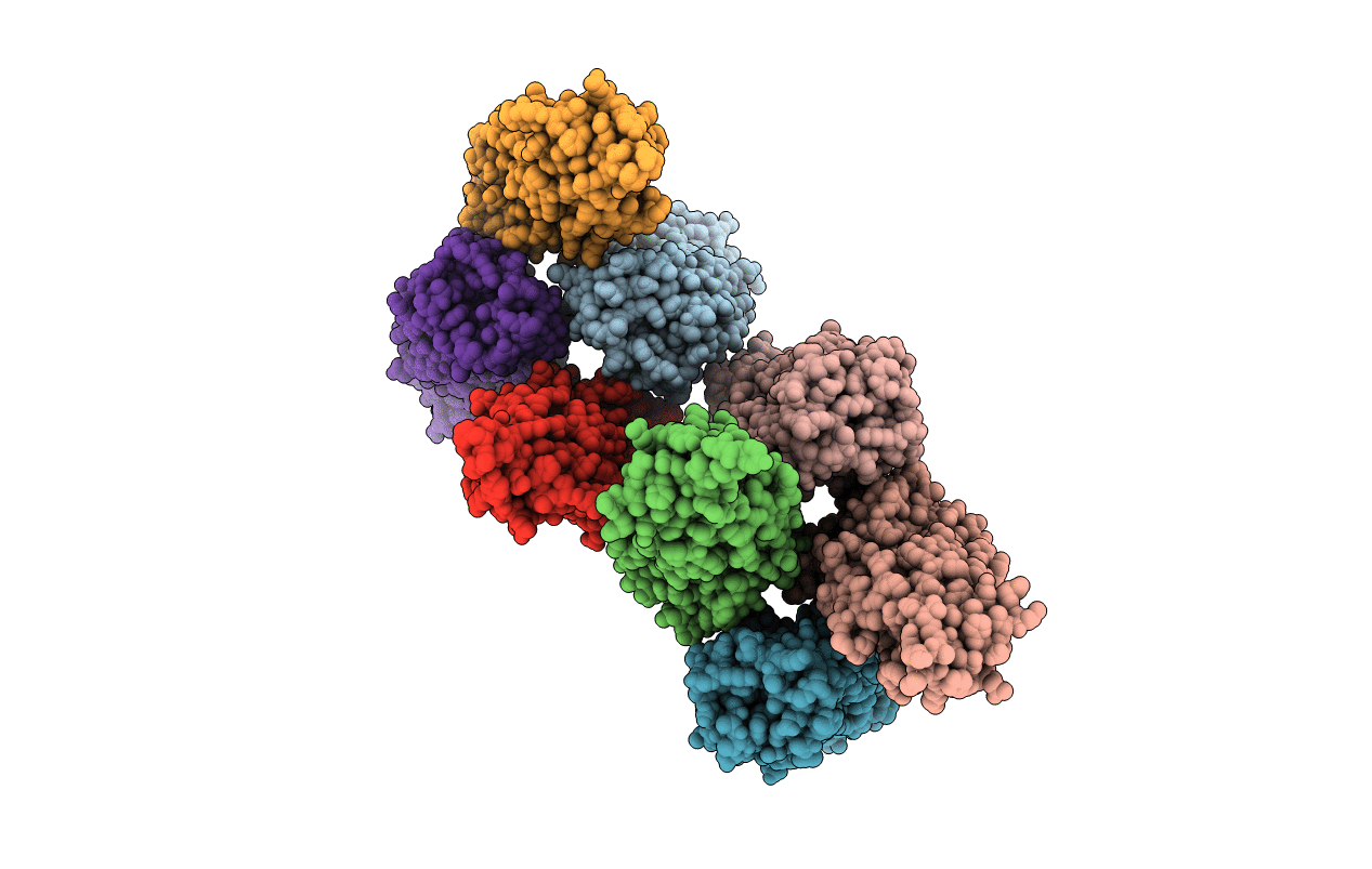
Title:
STRUCTURE OF THE PANTON-VALENTINE LEUCOCIDIN S COMPONENT FROM STAPHYLOCOCCUS AUREUS
Biological Source:
Source Organism(s):
Staphylococcus phage PVL (Taxon ID: 71366)
Method Details:
Experimental Method:
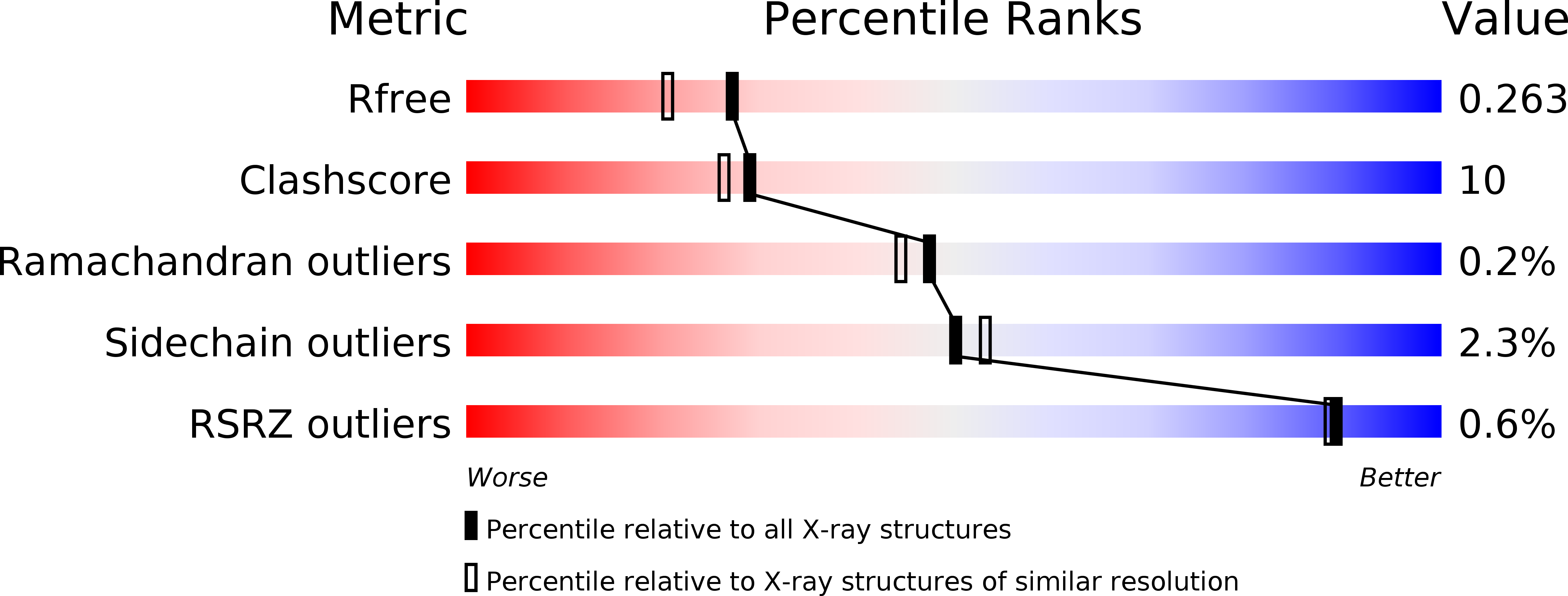
Resolution:
2.00 Å
R-Value Free:
0.24
R-Value Work:
0.20
Space Group:
P 43