1T5L image
Deposition Date 2004-05-04
Release Date 2004-06-22
Last Version Date 2024-02-14
Entry Detail
PDB ID:
1T5L
Title:
Crystal structure of the DNA repair protein UvrB point mutant Y96A revealing a novel fold for domain 2
Biological Source:
Source Organism:
Host Organism:
Method Details:
Experimental Method:
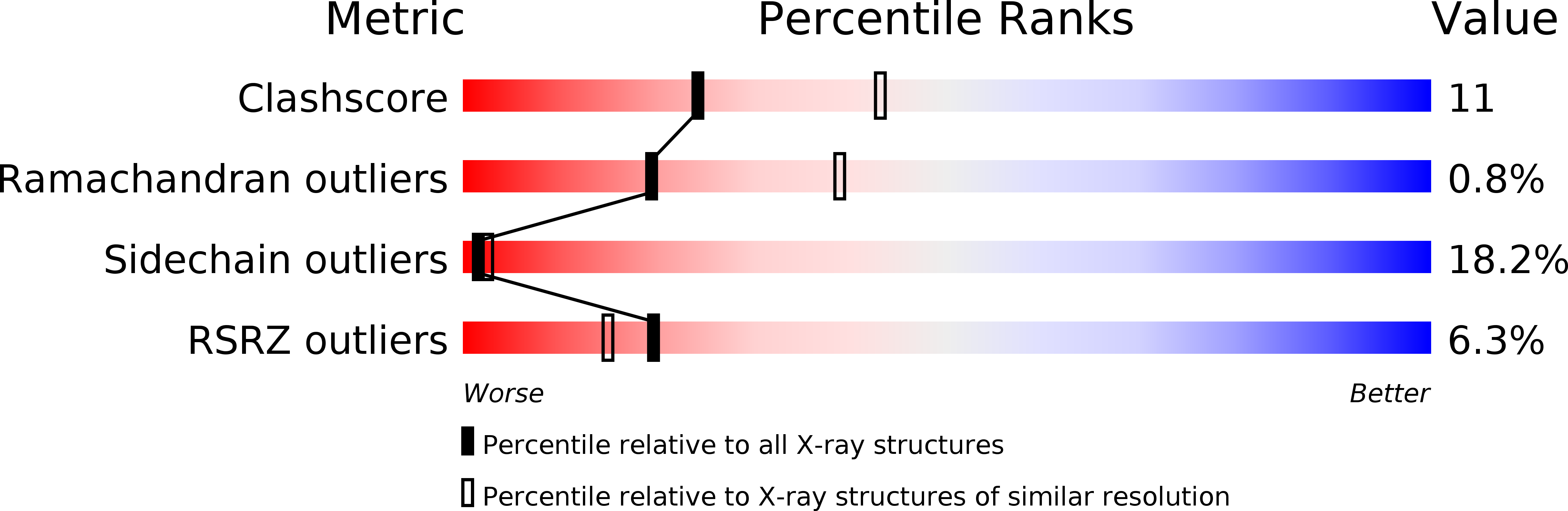
Resolution:
2.60 Å
R-Value Free:
0.28
R-Value Work:
0.22
R-Value Observed:
0.23
Space Group:
P 32 2 1
Macromolecular Entities
Polymer Type:polypeptide(L)
Molecule:UvrABC system protein B
Gene (Uniprot):uvrB
Mutations:Y96A
Chain IDs:A, B
Chain Length:658
Number of Molecules:2
Biological Source:Bacillus caldotenax
Ligand Molecules
Primary Citation
Interactions between UvrA and UvrB: the role of UvrB's domain 2 in nucleotide excision repair
Embo J. 23 2498 2509 (2004)
PMID: 15192705 DOI: 10.1038/sj.emboj.7600263

Abstact

Nucleotide excision repair (NER) is a highly conserved DNA repair mechanism present in all kingdoms of life. UvrB is a central component of the bacterial NER system, participating in damage recognition, strand excision and repair synthesis. None of the three presently available crystal structures of UvrB has defined the structure of domain 2, which is critical for the interaction with UvrA. We have solved the crystal structure of the UvrB Y96A variant, which reveals a new fold for domain 2 and identifies highly conserved residues located on its surface. These residues are restricted to the face of UvrB important for DNA binding and may be critical for the interaction of UvrB with UvrA. We have mutated these residues to study their role in the incision reaction, formation of the pre-incision complex, destabilization of short duplex regions in DNA, binding to UvrA and ATP hydrolysis. Based on the structural and biochemical data, we conclude that domain 2 is required for a productive UvrA-UvrB interaction, which is a pre-requisite for all subsequent steps in nucleotide excision repair.

Legend

Protein

Chemical

Disease

Primary Citation of related structures