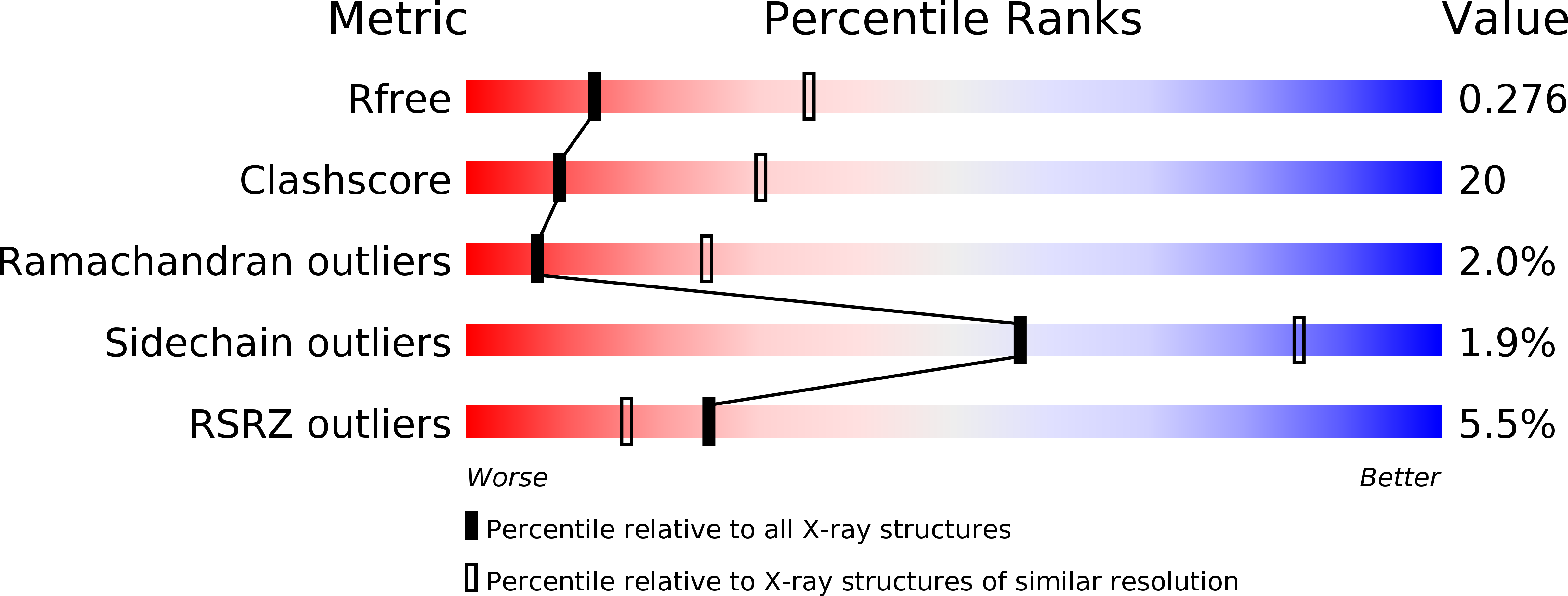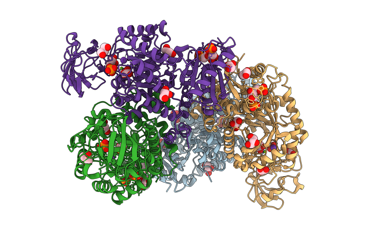
1T5A image
Deposition Date 2004-05-03
Release Date 2005-07-12
Last Version Date 2023-09-20
Entry Detail
PDB ID:
1T5A
Keywords:
Title:
Human Pyruvate Kinase M2
Biological Source:
Source Organism:
Homo sapiens (Taxon ID: 9606)
Host Organism:
Method Details:
Experimental Method:
Resolution:
2.80 Å
R-Value Free:
0.27
R-Value Work:
0.23
Space Group:
P 21 21 21
Macromolecular Entities
Polymer Type:polypeptide(L)
Molecule:Pyruvate kinase, M2 isozyme
Gene (Uniprot):PKM
Chain IDs:A, B, C, D
Chain Length:567
Number of Molecules:4
Biological Source:Homo sapiens
Primary Citation
Structural basis for tumor pyruvate kinase M2 allosteric regulation and catalysis.
Biochemistry 44 9417 9429 (2005)
PMID: 15996096 DOI: 10.1021/bi0474923

Abstact

Four isozymes of pyruvate kinase are differentially expressed in human tissue. Human pyruvate kinase isozyme M2 (hPKM2) is expressed in early fetal tissues and is progressively replaced by the other three isozymes, M1, R, and L, immediately after birth. In most cancer cells, hPKM2 is once again expressed to promote tumor cell proliferation. Because of its almost ubiquitous presence in cancer cells, hPKM2 has been designated as tumor specific PK-M2, and its presence in human plasma is currently being used as a molecular marker for the diagnosis of various cancers. The X-ray structure of human hPKM2 complexed with Mg(2+), K(+), the inhibitor oxalate, and the allosteric activator fructose 1,6-bisphosphate (FBP) has been determined to a resolution of 2.82 A. The active site of hPKM2 is in a partially closed conformation most likely resulting from a ligand-induced domain closure promoted by the binding of FBP. In all four subunits of the enzyme tetramer, a conserved water molecule is observed on the 2-si face of the prospective enolate and supports the hypothesis that a proton-relay system is acting as the proton donor of the reaction (1). Significant structural differences among the human M2, rabbit muscle M1, and the human R isozymes are observed, especially in the orientation of the FBP-activating loop, which is in a closed conformation when FBP is bound. The structural differences observed between the PK isozymes could potentially be exploited as unique structural templates for the design of allosteric drugs against the disease states associated with the various PK isozymes, especially cancer and nonspherocytic hemolytic anemia.

Legend

Protein

Chemical

Disease

Primary Citation of related structures