1T4W image
Deposition Date 2004-04-30
Release Date 2004-07-20
Last Version Date 2024-02-14
Entry Detail
PDB ID:
1T4W
Keywords:
Title:
Structural Differences in the DNA Binding Domains of Human p53 and its C. elegans Ortholog Cep-1: Structure of C. elegans Cep-1
Biological Source:
Source Organism:
Host Organism:
Method Details:
Experimental Method:
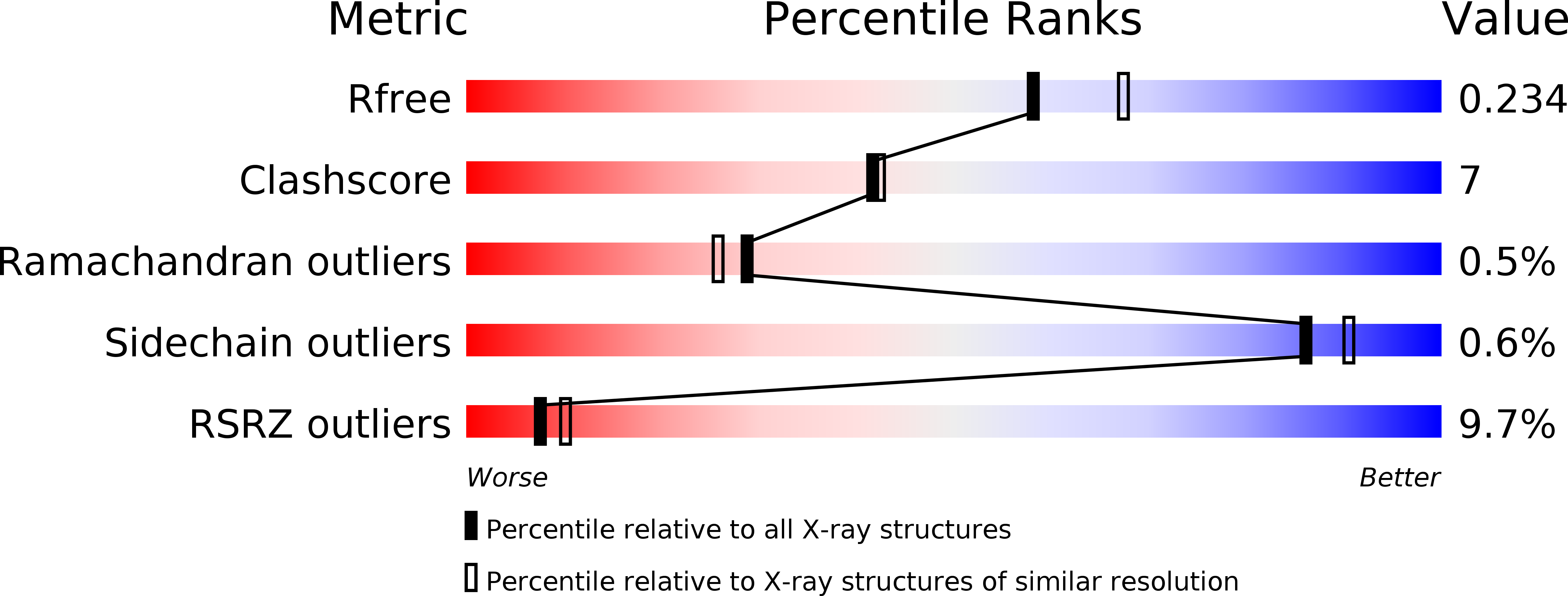
Resolution:
2.10 Å
R-Value Free:
0.23
R-Value Work:
0.17
R-Value Observed:
0.22
Space Group:
P 21 21 21
Macromolecular Entities
Polymer Type:polypeptide(L)
Molecule:C.Elegans p53 tumor suppressor-like transcription factor
Gene (Uniprot):cep-1
Chain IDs:A
Chain Length:196
Number of Molecules:1
Biological Source:Caenorhabditis elegans
Ligand Molecules
Primary Citation
Structural Differences in the DNA Binding Domains of Human p53 and Its C. elegans Ortholog Cep-1.
Structure 12 1237 1243 (2004)
PMID: 15242600 DOI: 10.1016/j.str.2004.05.007

Abstact

The DNA binding domains of human p53 and Cep-1, its C. elegans ortholog, recognize essentially identical DNA sequences despite poor sequence similarity. We solved the three-dimensional structure of the Cep-1 DNA binding domain in the absence of DNA and compared it to that of human p53. The two domains have similar overall folds. However, three loops, involved in DNA and Zn binding in human p53, contain small alpha helices in Cep-1. The alpha helix in loop L3 of Cep-1 orients the side chains of two conserved arginines toward DNA; in human p53, both arginines are mutation hotspots, but only one contacts DNA. The alpha helix in loop L1 of Cep-1 repositions the entire loop, making it unlikely for residues of this loop to contact bases in the major groove of DNA, as occurs in human p53. Thus, during evolution there have been considerable changes in the structure of the p53 DNA binding domain.

Legend

Protein

Chemical

Disease

Primary Citation of related structures