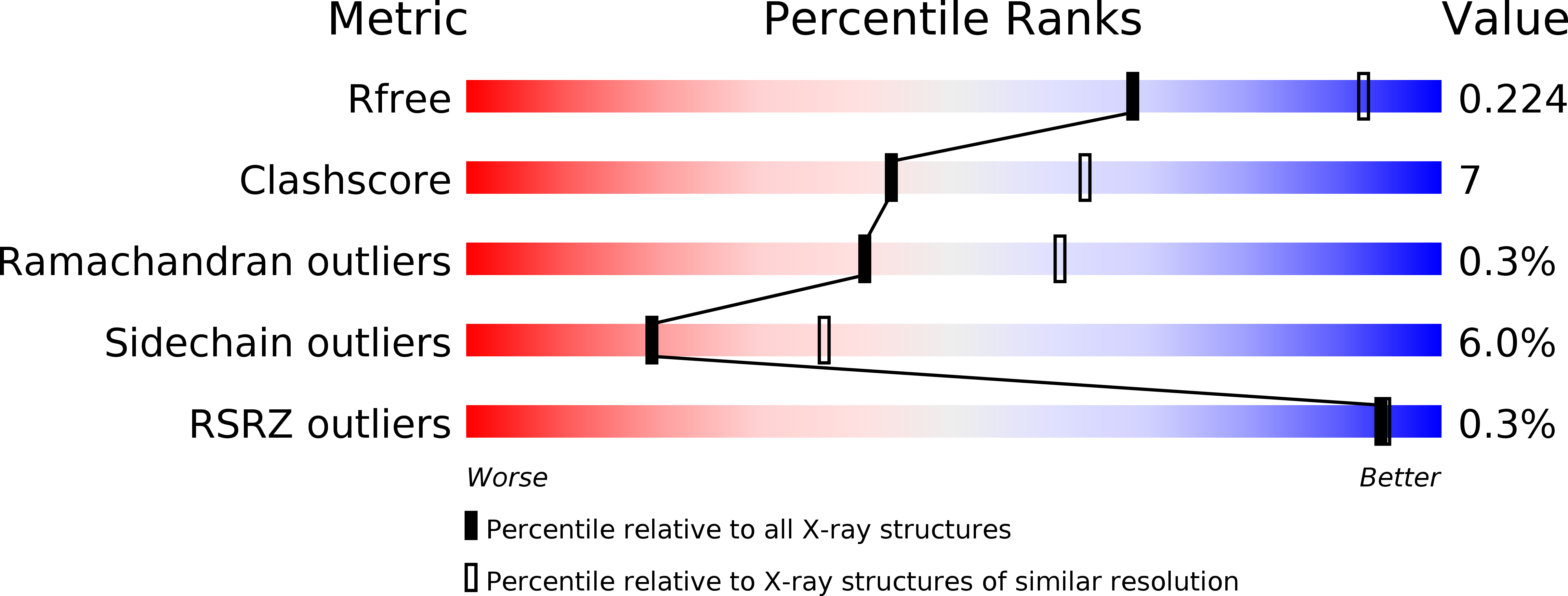
1T47 image
Deposition Date 2004-04-28
Release Date 2004-06-15
Last Version Date 2024-02-14
Entry Detail
PDB ID:
1T47
Keywords:
Title:
Structure of fe2-HPPD bound to NTBC
Biological Source:
Source Organism:
Method Details:
Experimental Method:
Resolution:
2.50 Å
R-Value Free:
0.23
R-Value Work:
0.17
R-Value Observed:
0.17
Space Group:
P 21 21 2
Macromolecular Entities
Structures with similar UniProt ID
Protein Blast
Polymer Type:polypeptide(L)
Molecule:4-hydroxyphenylpyruvate dioxygenase
Gene (Uniprot):hpd
Chain IDs:A, B
Chain Length:381
Number of Molecules:2
Biological Source:Streptomyces avermitilis
Primary Citation
Structure of the Ferrous Form of (4-Hydroxyphenyl)pyruvate Dioxygenase from Streptomyces avermitilis in Complex with the Therapeutic Herbicide, NTBC
Biochemistry 43 6370 6377 (2004)
PMID: 15157070 DOI: 10.1021/bi049317s

Abstact

Di- and triketone inhibitors of (4-hydroxyphenyl)pyruvate dioxygenase (HPPD) are both effective herbicides and therapeutics. The inhibitory activity is used to halt the production of lipophilic redox cofactors in plants and also in humans to prevent accumulation of toxic metabolic byproducts that arise from specific inborn defects of tyrosine catabolism. The three-dimensional structure of the Fe(II) form of HPPD from Streptomyces avermitilis in complex with the inhibitor 2-[2-nitro-4-(triflouromethyl)benzoyl]-1,3-cyclohexanedione (NTBC) has been determined at a resolution of 2.5 A. NTBC coordinates to the active site metal ion, located at the bottom of a wide solvent-accessible cavity in the C-terminal domain of the protein. The iron is liganded in a predominantly five-coordinate, distorted square-pyramidal arrangement in which Glu349, His187, and His270 are protein-derived ligands and two other ligands are from the 5' and 7' oxygens of NTBC. There is a low-occupancy water molecule in the sixth coordination site in one of the protomers. The distance to His270 is unusually long at 2.5 A, and its orientation is somewhat distorted from ideal ligand geometry to within 2.8 A of the inhibitor nitro group. In contrast to the tetrameric quartenary structure observed for HPPD from other bacterial sources, the asymmetric unit is composed of two weakly associated protomers with a buried surface area of 1266 A(2) and a total of 12 hydrogen-bonding and no electrostatic interactions. The overall tertiary structure is similar to that of HPPD from Pseudomonas fluorescens (Serre et al., (1999) Structure 7, 977-988), although the position of the C-terminal alpha-helix is dramatically shifted. This C-terminal alpha-helix provides Phe364, which in combination with Phe336 sandwiches the phenyl ring of the bound NTBC; no other significant hydrogen-bonding or charge-pairing interactions are observed. Moreover, the structure reveals that, with the exception of Val189, NTBC makes contacts to only fully conserved amino acids. The combination of bidentate metal-ion coordination and pi-stacked aromatic rings is suggestive of a binding mode for the substrate and/or a transition state, which may be the origin of the exceedingly high affinity these inhibitors have for HPPD.

Legend

Protein

Chemical

Disease

Primary Citation of related structures