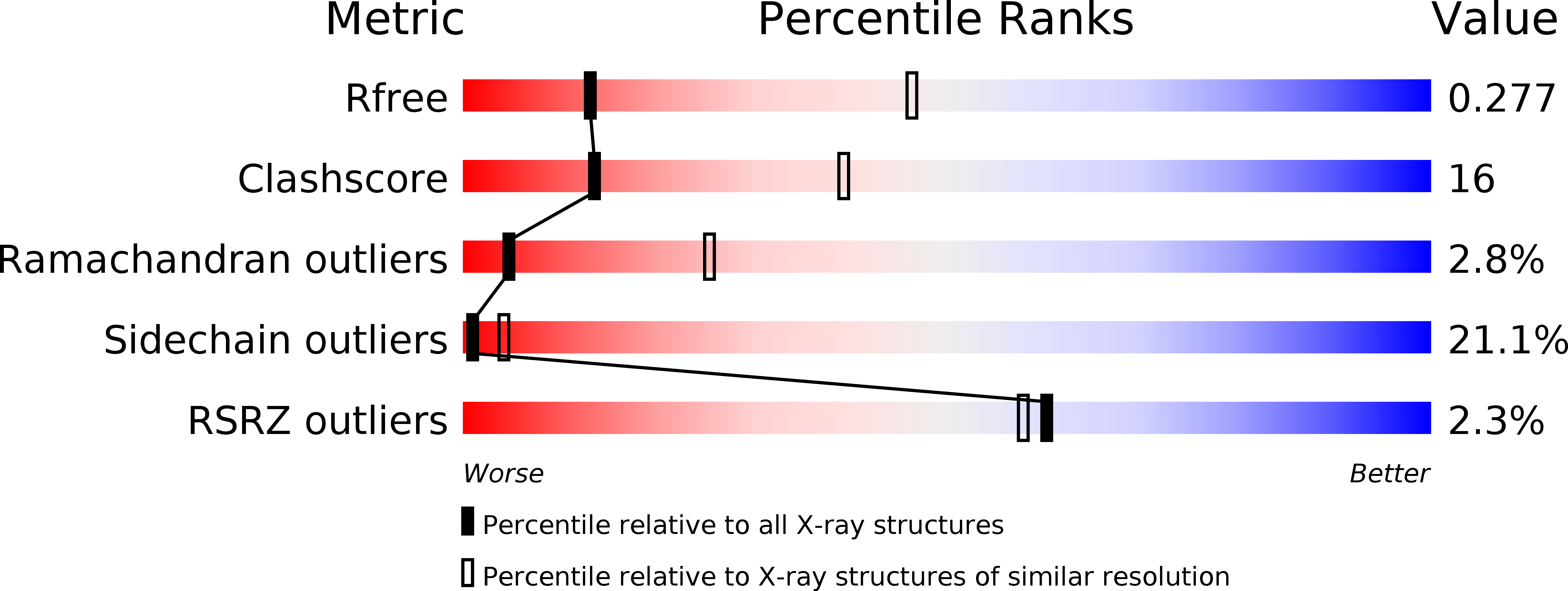
1T3E image
Deposition Date 2004-04-26
Release Date 2004-07-27
Last Version Date 2024-04-03
Entry Detail
PDB ID:
1T3E
Title:
Structural basis of dynamic glycine receptor clustering
Biological Source:
Source Organism:
Host Organism:
Method Details:
Experimental Method:
Resolution:
3.25 Å
R-Value Free:
0.30
R-Value Work:
0.24
R-Value Observed:
0.24
Space Group:
P 61
Macromolecular Entities
Polymer Type:polypeptide(L)
Molecule:Gephyrin
Gene (Uniprot):Gphn
Chain IDs:A, B
Chain Length:421
Number of Molecules:2
Biological Source:Rattus norvegicus
Polymer Type:polypeptide(L)
Molecule:49-mer fragment of Glycine receptor beta chain
Gene (Uniprot):Glrb
Chain IDs:C (auth: P)
Chain Length:49
Number of Molecules:1
Biological Source:Rattus norvegicus
Ligand Molecules
Primary Citation
Structural basis of dynamic glycine receptor clustering by gephyrin
Embo J. 23 2510 2519 (2004)
PMID: 15201864 DOI: 10.1038/sj.emboj.7600256

Abstact

Gephyrin is a bi-functional modular protein involved in molybdenum cofactor biosynthesis and in postsynaptic clustering of inhibitory glycine receptors (GlyRs). Here, we show that full-length gephyrin is a trimer and that its proteolysis in vitro causes the spontaneous dimerization of its C-terminal region (gephyrin-E), which binds a GlyR beta-subunit-derived peptide with high and low affinity. The crystal structure of the tetra-domain gephyrin-E in complex with the beta-peptide bound to domain IV indicates how membrane-embedded GlyRs may interact with subsynaptic gephyrin. In vitro, trimeric full-length gephyrin forms a network upon lowering the pH, and this process can be reversed to produce stable full-length dimeric gephyrin. Our data suggest a mechanism by which induced conformational transitions of trimeric gephyrin may generate a reversible postsynaptic scaffold for GlyR recruitment, which allows for dynamic receptor movement in and out of postsynaptic GlyR clusters, and thus for synaptic plasticity.

Legend

Protein

Chemical

Disease

Primary Citation of related structures