Deposition Date
2004-04-19
Release Date
2004-06-29
Last Version Date
2023-08-23
Entry Detail
PDB ID:
1T1V
Keywords:
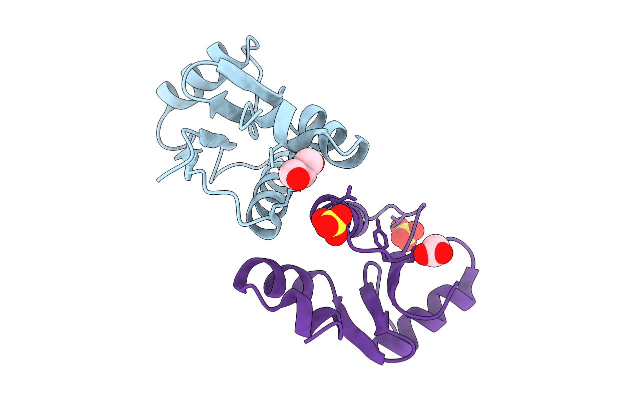
Title:
Crystal Structure of the Glutaredoxin-like Protein SH3BGRL3 at 1.6 A resolution
Biological Source:
Source Organism(s):
Mus musculus (Taxon ID: 10090)
Expression System(s):
Method Details:
Experimental Method:
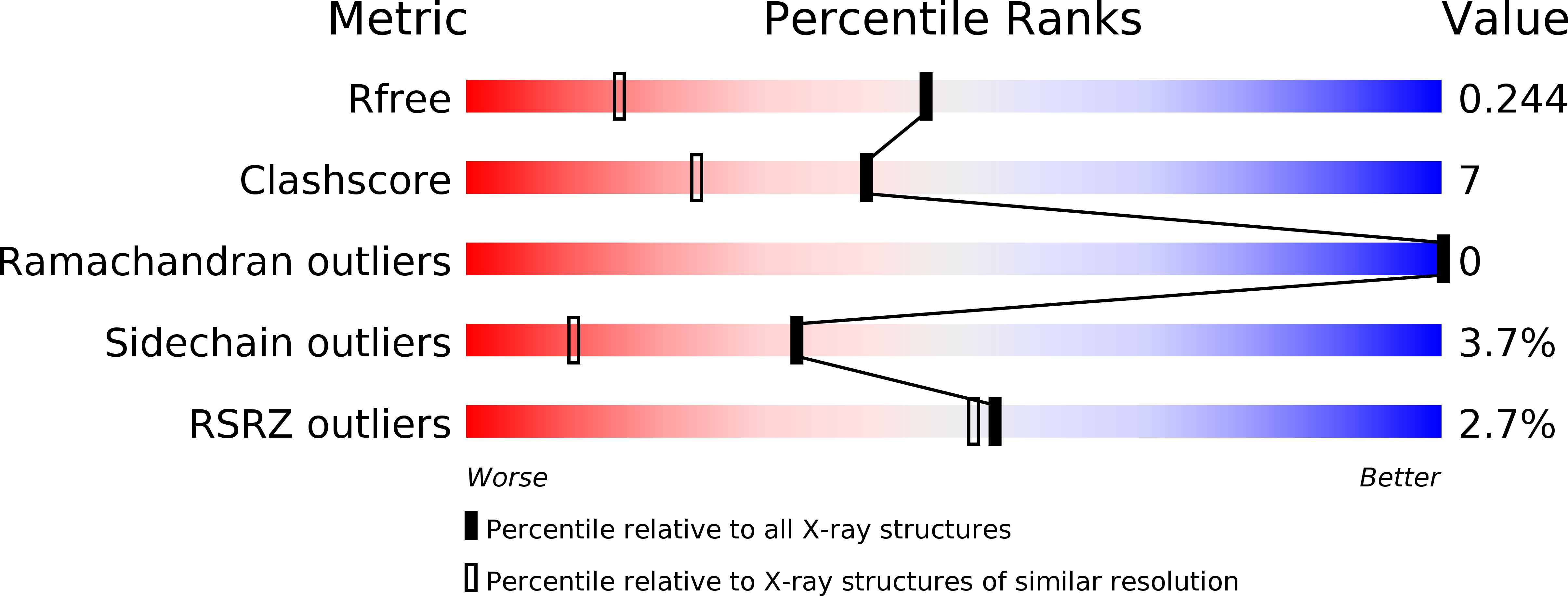
Resolution:
1.60 Å
R-Value Free:
0.24
R-Value Work:
0.19
R-Value Observed:
0.20
Space Group:
P 1 21 1