1T19 image
Entry Detail
PDB ID:
1T19
Keywords:
Title:
Early intermediate IE2 from time-resolved crystallography of the E46Q mutant of PYP
Biological Source:
Source Organism:
Host Organism:
PDB Version:
Deposition Date:
2004-04-15
Release Date:
2005-01-18
Method Details:
Experimental Method:
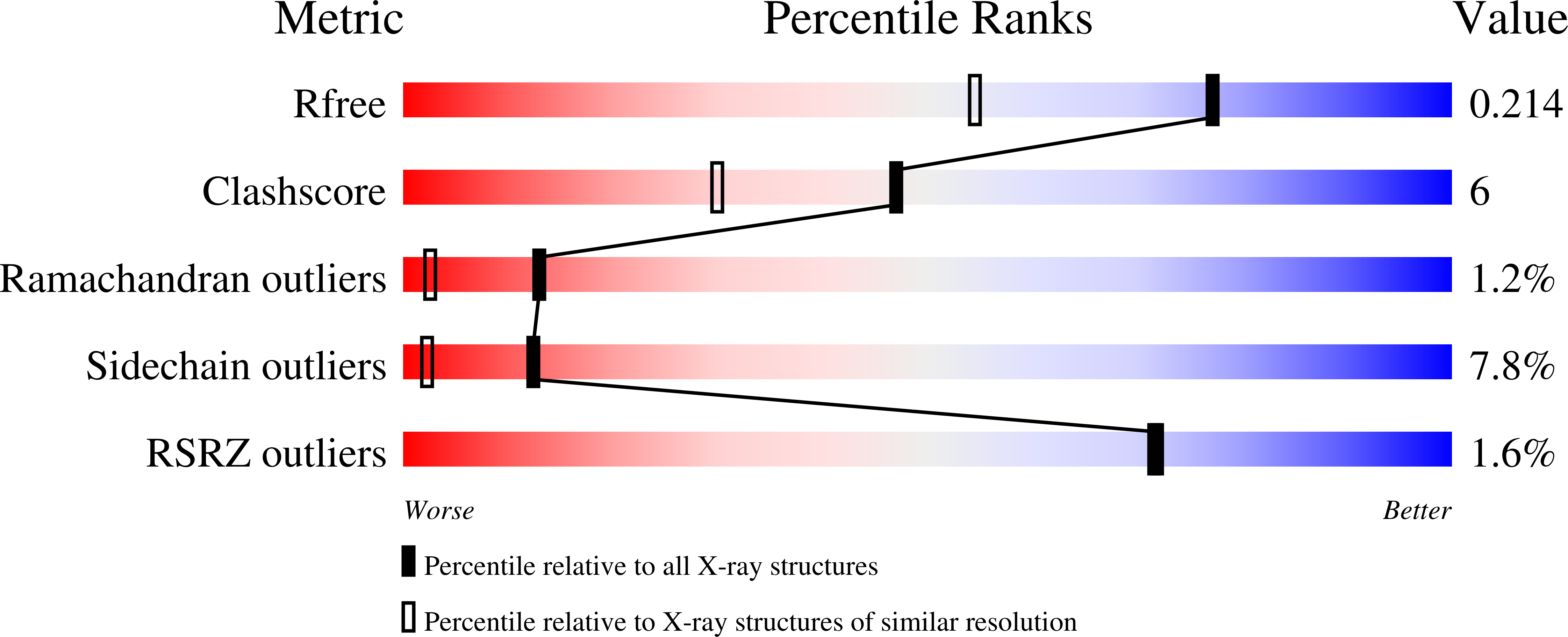
Resolution:
1.60 Å
R-Value Free:
0.21
R-Value Work:
0.20
R-Value Observed:
0.20
Space Group:
P 63
Macromolecular Entities
Polymer Type:polypeptide(L)
Description:Photoactive yellow protein
Mutations:E46Q mutant
Chain IDs:A
Chain Length:125
Number of Molecules:1
Biological Source:Halorhodospira halophila
Ligand Molecules
Primary Citation
A Structural Pathway for Signaling in the E46Q Mutant of Photoactive Yellow Protein
Structure 13 55 63 (2005)
PMID: 15642261 DOI: 10.1016/j.str.2004.10.016

Abstact

In the bacterial photoreceptor photoactive yellow protein (PYP), absorption of blue light by its chromophore leads to a conformational change in the protein associated with differential signaling activity, as it executes a reversible photocycle. Time-resolved Laue crystallography allows structural snapshots (as short as 150 ps) of high crystallographic resolution (approximately 1.6 A) to be taken of a protein as it functions. Here, we analyze by singular value decomposition a comprehensive time-resolved crystallographic data set of the E46Q mutant of PYP throughout the photocycle spanning 10 ns-100 ms. We identify and refine the structures of five distinct intermediates and provide a plausible chemical kinetic mechanism for their inter conversion. A clear structural progression is visible in these intermediates, in which a signal generated at the chromophore propagates through a distinct structural pathway of conserved residues and results in structural changes near the N terminus, over 20 A distant from the chromophore.

Legend

Protein

Chemical

Disease

Primary Citation of related structures