1T0J image
Deposition Date 2004-04-09
Release Date 2004-06-15
Last Version Date 2023-08-23
Entry Detail
PDB ID:
1T0J
Title:
Crystal structure of a complex between voltage-gated calcium channel beta2a subunit and a peptide of the alpha1c subunit
Biological Source:
Source Organism:
Rattus norvegicus (Taxon ID: 10116)
Homo sapiens (Taxon ID: 9606)
Host Organism:
Method Details:
Experimental Method:
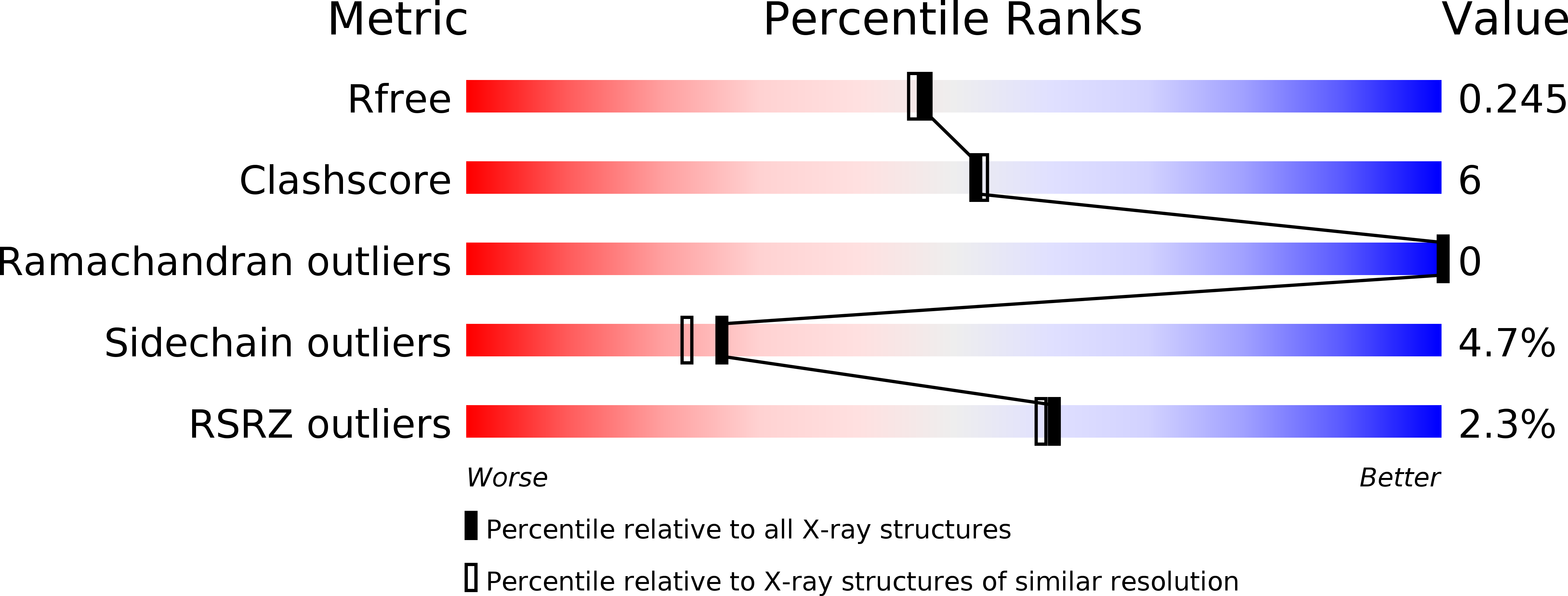
Resolution:
2.00 Å
R-Value Free:
0.24
R-Value Work:
0.19
R-Value Observed:
0.20
Space Group:
P 1
Macromolecular Entities
Structures with similar UniProt ID
Protein Blast
Polymer Type:polypeptide(L)
Molecule:voltage-gated calcium channel subunit beta2a
Gene (Uniprot):Cacnb2
Chain IDs:A
Chain Length:132
Number of Molecules:1
Biological Source:Rattus norvegicus
Structures with similar UniProt ID
Protein Blast
Polymer Type:polypeptide(L)
Molecule:voltage-gated calcium channel subunit beta2a
Gene (Uniprot):Cacnb2
Chain IDs:B
Chain Length:224
Number of Molecules:1
Biological Source:Rattus norvegicus
Structures with similar UniProt ID
Protein Blast
Polymer Type:polypeptide(L)
Molecule:Voltage-dependent L-type calcium channel alpha-1C subunit
Gene (Uniprot):CACNA1C
Chain IDs:C
Chain Length:20
Number of Molecules:1
Biological Source:Homo sapiens
Ligand Molecules
Primary Citation
Structure of a complex between a voltage-gated calcium channel beta-subunit and an alpha-subunit domain.
Nature 429 671 675 (2004)
PMID: 15141227 DOI: 10.1038/nature02588

Abstact

Voltage-gated calcium channels (Ca(V)s) govern muscle contraction, hormone and neurotransmitter release, neuronal migration, activation of calcium-dependent signalling cascades, and synaptic input integration. An essential Ca(V) intracellular protein, the beta-subunit (Ca(V)beta), binds a conserved domain (the alpha-interaction domain, AID) between transmembrane domains I and II of the pore-forming alpha(1) subunit and profoundly affects multiple channel properties such as voltage-dependent activation, inactivation rates, G-protein modulation, drug sensitivity and cell surface expression. Here, we report the high-resolution crystal structures of the Ca(V)beta2a conserved core, alone and in complex with the AID. Previous work suggested that a conserved region, the beta-interaction domain (BID), formed the AID-binding site; however, this region is largely buried in the Ca(V)beta core and is unavailable for protein-protein interactions. The structure of the AID-Ca(V)beta2a complex shows instead that Ca(V)beta2a engages the AID through an extensive, conserved hydrophobic cleft (named the alpha-binding pocket, ABP). The ABP-AID interaction positions one end of the Ca(V)beta near the intracellular end of a pore-lining segment, called IS6, that has a critical role in Ca(V) inactivation. Together, these data suggest that Ca(V)betas influence Ca(V) gating by direct modulation of IS6 movement within the channel pore.

Legend

Protein

Chemical

Disease

Primary Citation of related structures