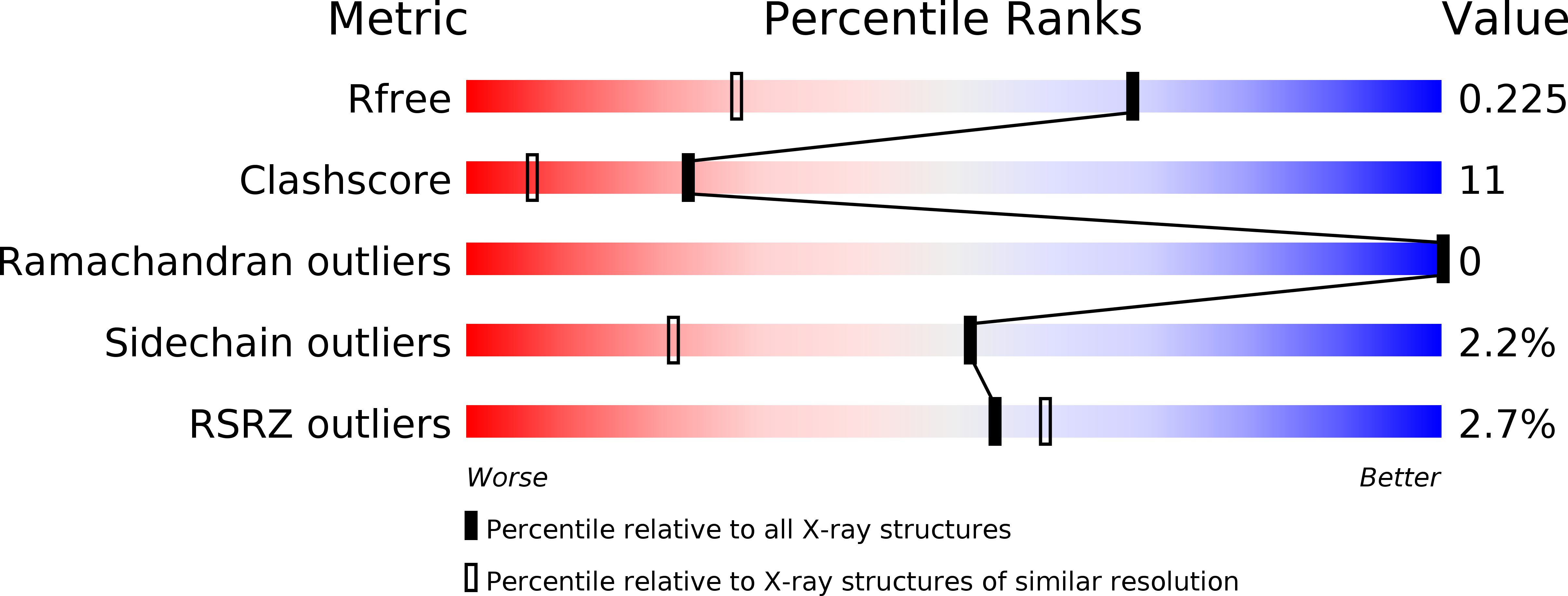
1T00 image
Deposition Date 2004-04-07
Release Date 2005-01-25
Last Version Date 2024-02-14
Entry Detail
PDB ID:
1T00
Title:
The structure of thioredoxin from S. coelicolor
Biological Source:
Source Organism:
Method Details:
Experimental Method:
Resolution:
1.51 Å
R-Value Free:
0.21
R-Value Work:
0.17
R-Value Observed:
0.17
Space Group:
P 21 21 2
Macromolecular Entities
Structures with similar UniProt ID
Protein Blast
Polymer Type:polypeptide(L)
Molecule:Thioredoxin
Gene (Uniprot):trxA
Chain IDs:A
Chain Length:112
Number of Molecules:1
Biological Source:Streptomyces coelicolor
Primary Citation
Expression, purification and X-ray crystallographic analysis of thioredoxin from Streptomyces coelicolor.
Acta Crystallogr.,Sect.F 61 164 168 (2005)
PMID: 16510983 DOI: 10.1107/S1744309104032993

Abstact

Thioredoxins are ubiquitous proteins that serve as reducing agents and general protein disulfide reductases. In turn, they are reduced by electrons obtained from the NADPH-containing thioredoxin reductase. Thioredoxins have been isolated and characterized from a large number of organisms. The Gram-positive bacterium Streptomyces coelicolor contains three thioredoxins that are involved in unknown biological processes. trxA from S. coelicolor was cloned and expressed in Escherichia coli and the protein purified and crystallized using the hanging-drop method of vapour diffusion. The crystal structure of thioredoxin A has been determined at 1.5 A resolution using a synchrotron-radiation source. The protein reveals a thioredoxin-like fold with a typical CXXC active site. The crystal exhibits the symmetry of space group P2(1)2(1)2, with unit-cell parameters a = 43.6, b = 71.8, c = 33.2 A.

Legend

Protein

Chemical

Disease

Primary Citation of related structures