
Deposition Date
2004-04-06
Release Date
2004-06-01
Last Version Date
2024-11-20
Entry Detail
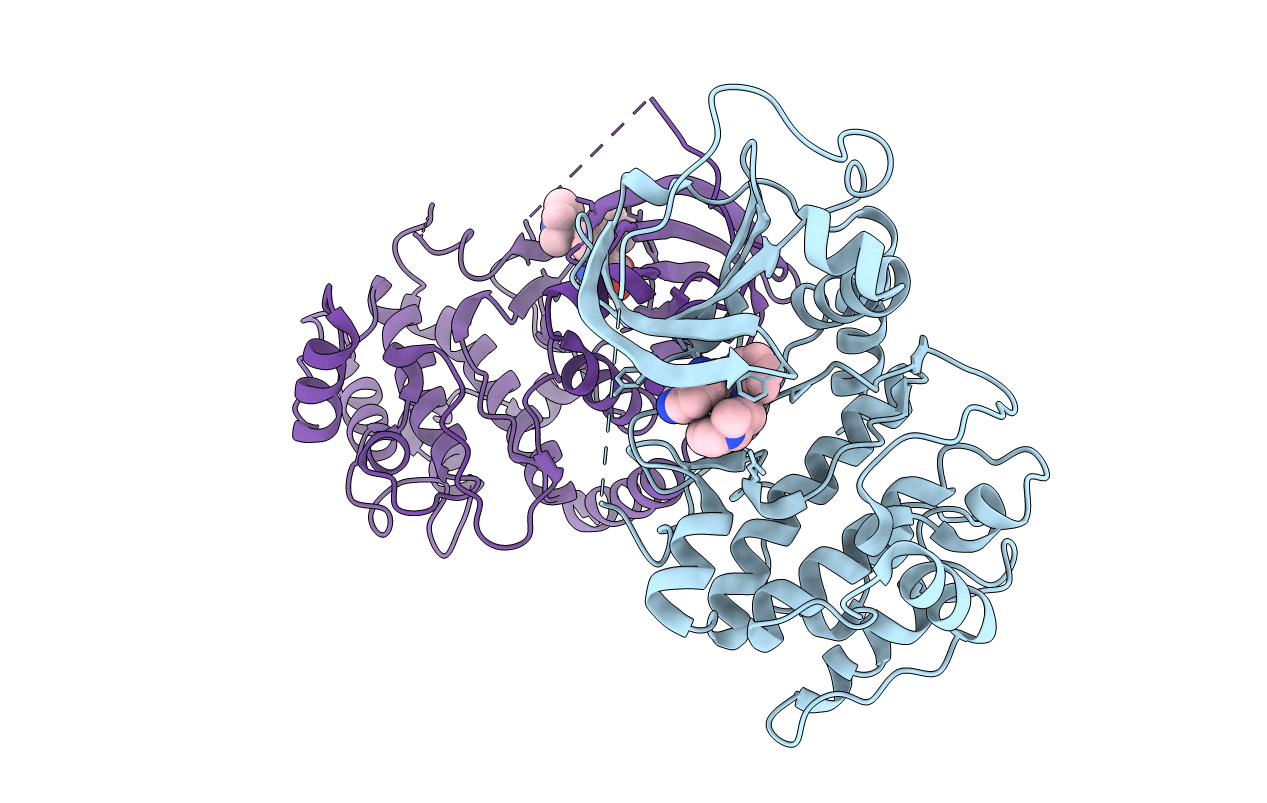
PDB ID:
1SZM
Keywords:
Title:
DUAL BINDING MODE OF BISINDOLYLMALEIMIDE 2 TO PROTEIN KINASE A (PKA)
Biological Source:
Source Organism(s):
Bos taurus (Taxon ID: 9913)
Expression System(s):
Method Details:
Experimental Method:
Resolution:
2.50 Å
R-Value Free:
0.28
R-Value Work:
0.23
R-Value Observed:
0.23
Space Group:
P 21 21 21
