
Deposition Date
2004-04-02
Release Date
2004-07-13
Last Version Date
2024-02-14
Entry Detail
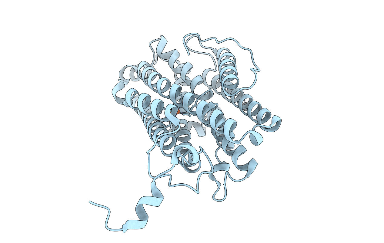
PDB ID:
1SYY
Keywords:
Title:
Crystal structure of the R2 subunit of ribonucleotide reductase from Chlamydia trachomatis
Biological Source:
Source Organism(s):
Chlamydia trachomatis (Taxon ID: 813)
Expression System(s):
Method Details:
Experimental Method:
Resolution:
1.70 Å
R-Value Free:
0.19
R-Value Work:
0.15
R-Value Observed:
0.15
Space Group:
P 43 21 2
