Deposition Date
2004-03-30
Release Date
2004-06-29
Last Version Date
2023-08-23
Entry Detail
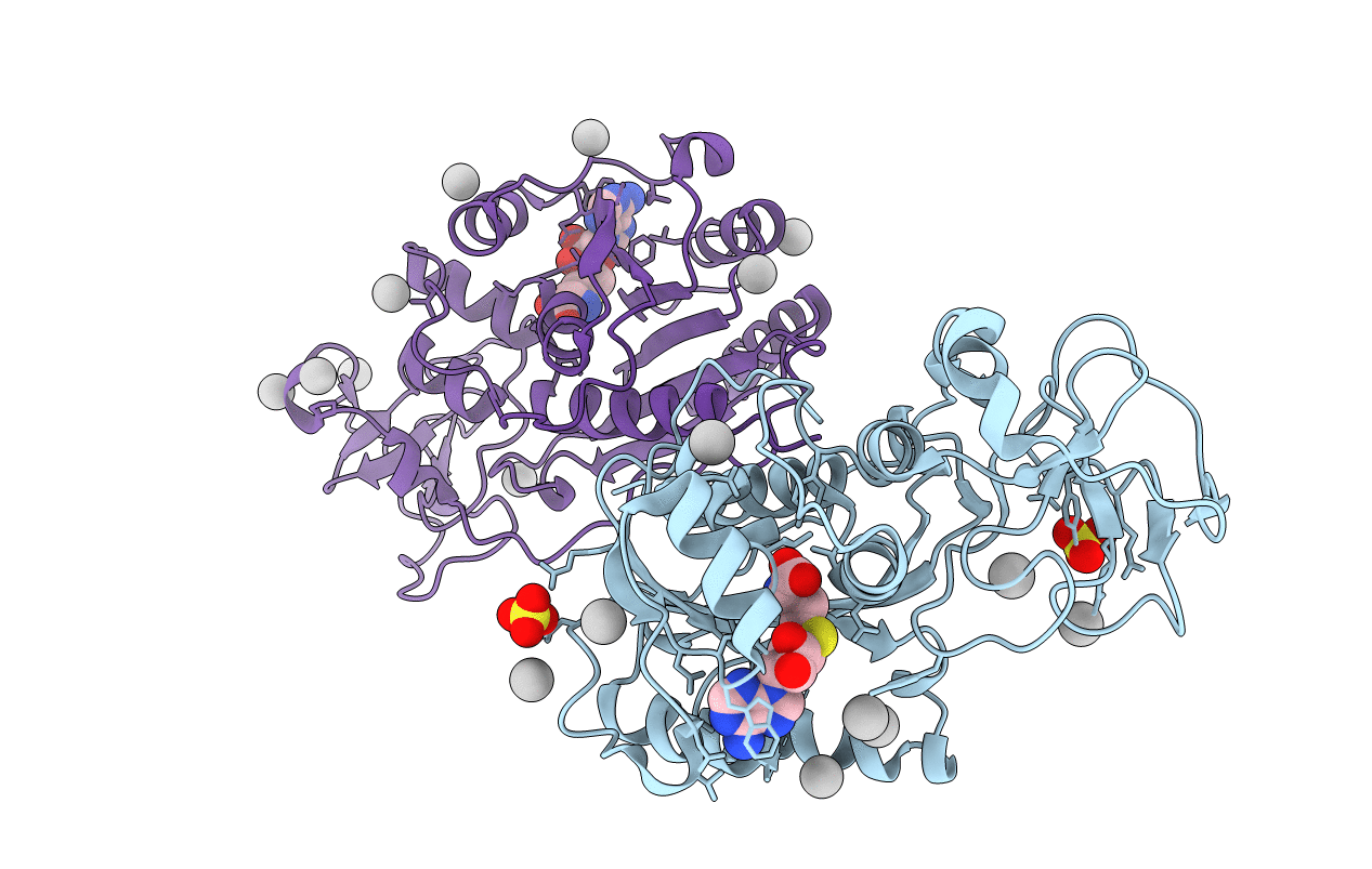
PDB ID:
1SVU
Keywords:
Title:
Structure of the Q237W mutant of HhaI DNA methyltransferase: an insight into protein-protein interactions
Biological Source:
Source Organism(s):
Haemophilus haemolyticus (Taxon ID: 726)
Expression System(s):
Method Details:
Experimental Method:
Resolution:
2.66 Å
R-Value Free:
0.26
R-Value Work:
0.18
R-Value Observed:
0.18
Space Group:
C 1 2 1