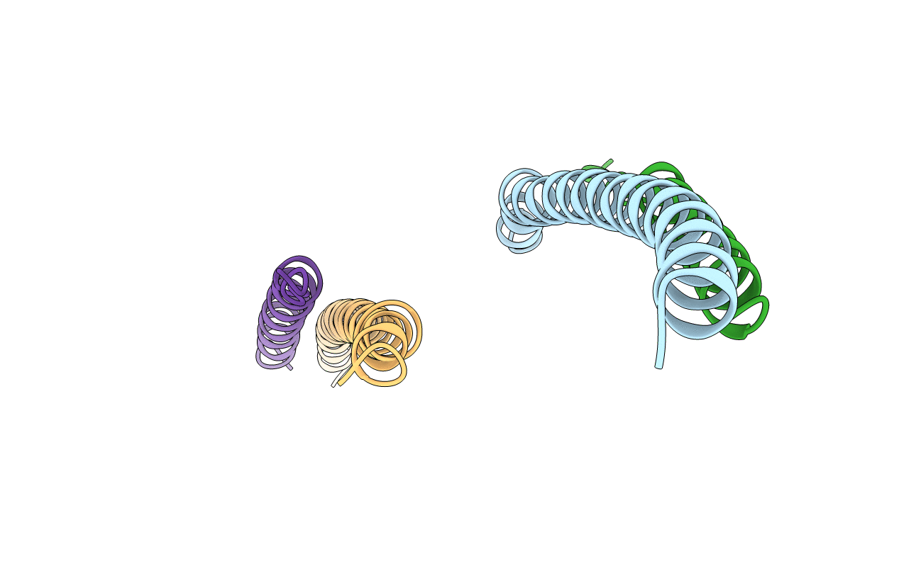
1SVF image
Deposition Date 1999-02-27
Release Date 1999-03-26
Last Version Date 2023-12-27
Entry Detail
PDB ID:
1SVF
Keywords:
Title:
PARAMYXOVIRUS SV5 FUSION PROTEIN CORE
Biological Source:
Source Organism:
Host Organism:
Method Details:
Experimental Method:
Resolution:
1.40 Å
R-Value Free:
0.20
R-Value Work:
0.18
Space Group:
P 3
Macromolecular Entities
Polymer Type:polypeptide(L)
Molecule:PROTEIN (FUSION GLYCOPROTEIN)
Gene (Uniprot):F
Mutations:C185Y
Chain IDs:A, C
Chain Length:64
Number of Molecules:2
Biological Source:Simian virus 5 (strain W3)
Polymer Type:polypeptide(L)
Molecule:PROTEIN (FUSION GLYCOPROTEIN)
Gene (Uniprot):F
Chain IDs:B, D
Chain Length:38
Number of Molecules:2
Biological Source:Simian virus 5 (strain W3)
Ligand Molecules
Primary Citation
Structural basis for paramyxovirus-mediated membrane fusion.
Mol.Cell 3 309 319 (1999)
PMID: 10198633 DOI: 10.1016/S1097-2765(00)80458-X

Abstact

Paramyxoviruses are responsible for significant human mortality and disease worldwide, but the molecular mechanisms underlying their entry into host cells remain poorly understood. We have solved the crystal structure of a fragment of the simian parainfluenza virus 5 fusion protein (SV5 F), revealing a 96 A long coiled coil surrounded by three antiparallel helices. This structure places the fusion and transmembrane anchor of SV5 F in close proximity with a large intervening domain at the opposite end of the coiled coil. Six amino acids, potentially part of the fusion peptide, form a segment of the central coiled coil, suggesting that this structure extends into the membrane. Deletion mutants of SV5 F indicate that putative flexible tethers between the coiled coil and the viral membrane are dispensable for fusion. The lack of flexible tethers may couple a final conformational change in the F protein directly to the fusion of two bilayers.

Legend

Protein

Chemical

Disease

Primary Citation of related structures