Deposition Date
2004-03-26
Release Date
2004-07-20
Last Version Date
2023-08-23
Entry Detail
PDB ID:
1SUO
Keywords:
Title:
Structure of mammalian cytochrome P450 2B4 with bound 4-(4-chlorophenyl)imidazole
Biological Source:
Source Organism(s):
Oryctolagus cuniculus (Taxon ID: 9986)
Expression System(s):
Method Details:
Experimental Method:
Resolution:
1.90 Å
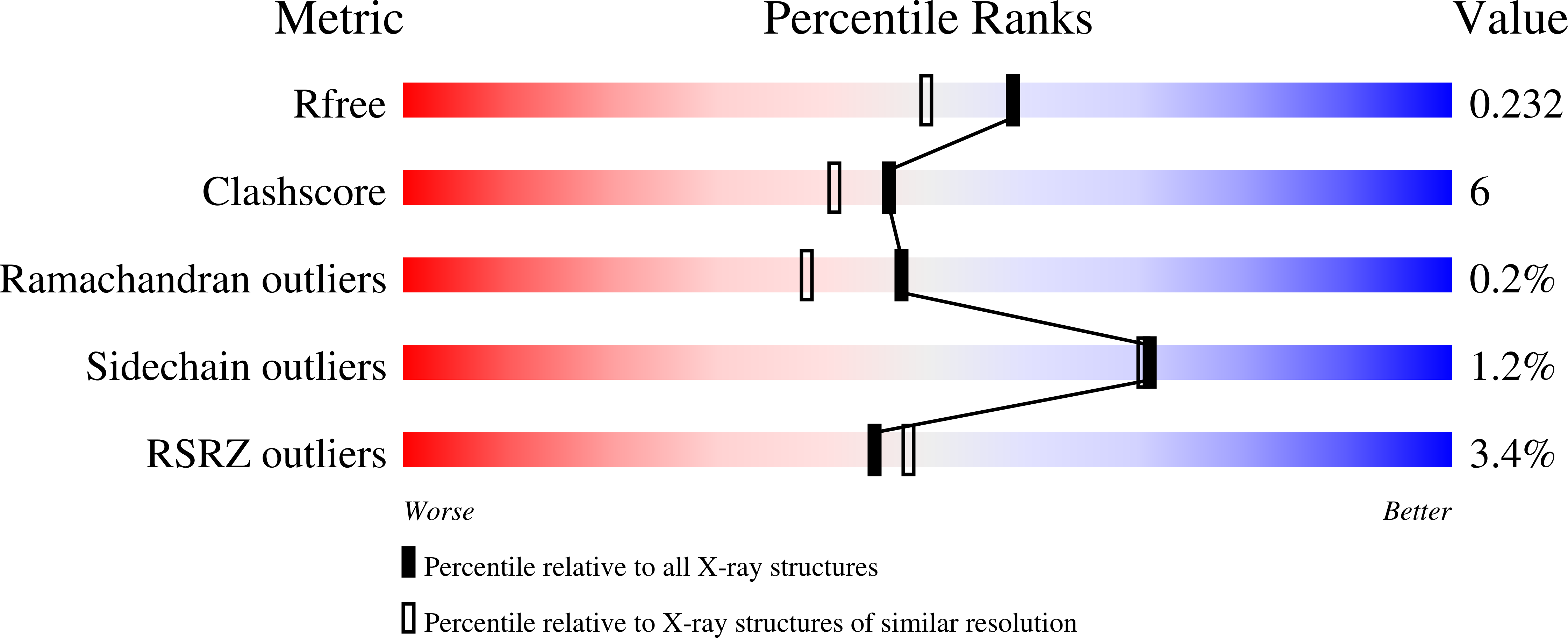
R-Value Free:
0.23
R-Value Work:
0.21
Space Group:
P 63 2 2