1ST7 image
Deposition Date 2004-03-25
Release Date 2005-03-01
Last Version Date 2024-05-22
Entry Detail
PDB ID:
1ST7
Title:
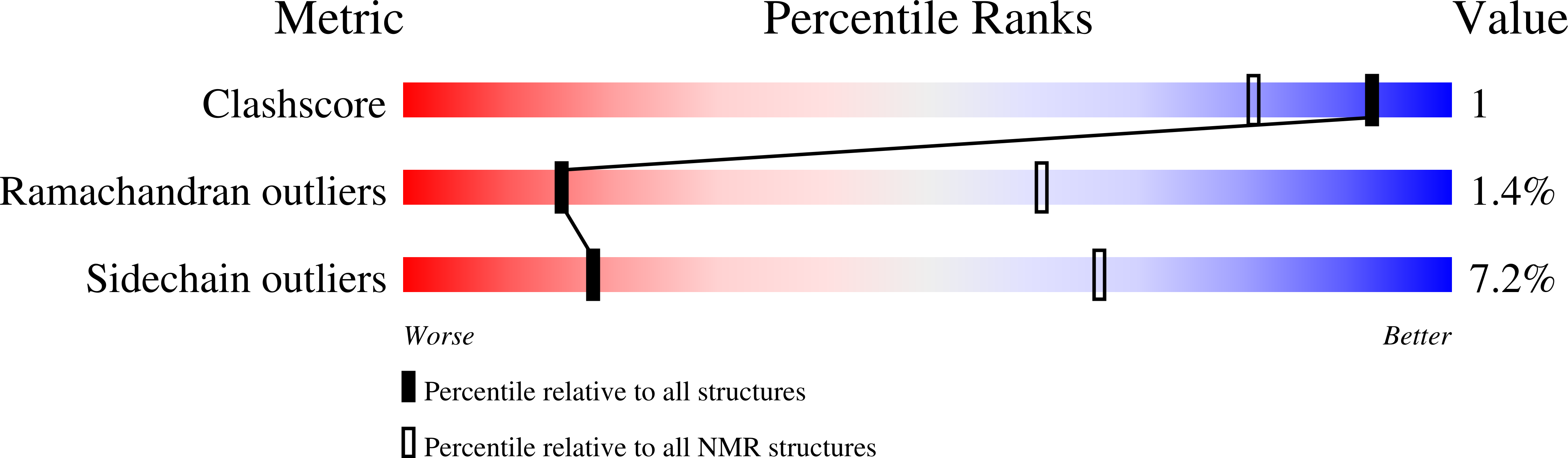
Solution structure of Acyl Coenzyme A Binding Protein from yeast
Biological Source:
Source Organism:
Host Organism:
Method Details:
Experimental Method:
Conformers Calculated:
20
Conformers Submitted:
20
Selection Criteria:
structures with acceptable covalent geometry,structures with the least restraint violations,structures with the lowest energy
Macromolecular Entities
Polymer Type:polypeptide(L)
Molecule:Acyl-CoA-binding protein
Gene (Uniprot):ACB1
Chain IDs:A
Chain Length:86
Number of Molecules:1
Biological Source:Saccharomyces cerevisiae
Ligand Molecules
Primary Citation
Different secondary structure elements as scaffolds for protein folding transition states of two homologous four-helix bundles
Proteins 59 80 90 (2005)
PMID: 15690348 DOI: 10.1002/prot.20340

Abstact

Comparison of the folding processes for homologue proteins can provide valuable information about details in the interactions leading to the formation of the folding transition state. Here the folding kinetics of 18 variants of yACBP and 3 variants of bACBP have been studied by Phi-value analysis. In combination with Phi-values from previous work, detailed insight into the transition states for folding of both yACBP and bACBP has been obtained. Of the 16 sequence positions that have been studied in both yACBP and bACBP, 5 (V12, I/L27, Y73, V77, and L80) have high Phi-values and appear to be important for the transition state formation in both homologues. Y31, A34, and A69 have high Phi-values only in yACBP, while F5, A9, and I74 have high Phi-values only in bACBP. Thus, additional interactions between helices A2 and A4 appear to be important for the transition state of yACBP, whereas additional interactions between helices A1 and A4 appear to be important for the transition state of bACBP. To examine whether these differences could be assigned to different packing of the residues in the native state, a solution structure of yACBP was determined by NMR. Small changes in the packing of the hydrophobic side-chains, which strengthen the interactions between helices A2 and A4, are observed in yACBP relative to bACBP. It is suggested that different structure elements serve as scaffolds for the folding of the 2 ACBP homologues.

Legend

Protein

Chemical

Disease

Primary Citation of related structures