1ST6 image
Deposition Date 2004-03-25
Release Date 2004-08-03
Last Version Date 2024-02-14
Entry Detail
PDB ID:
1ST6
Keywords:
Title:
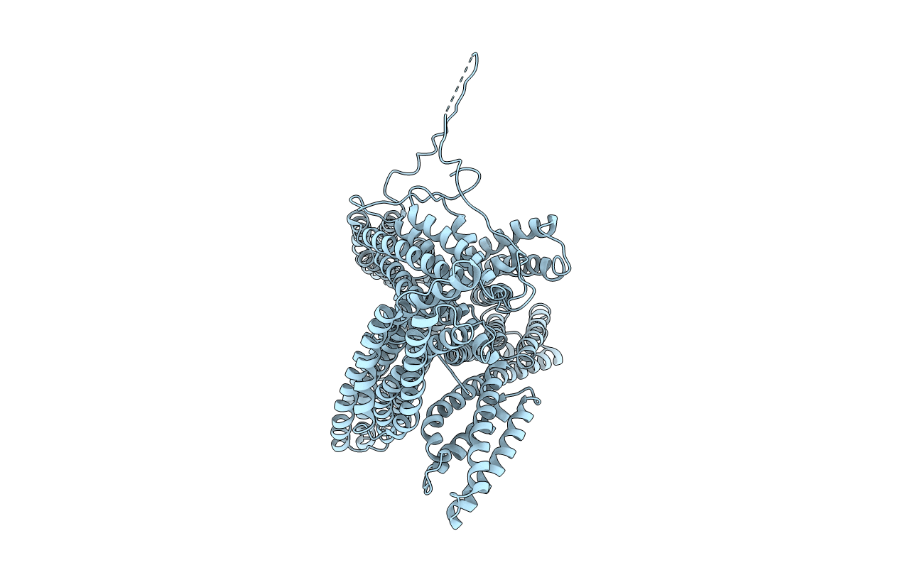
Crystal structure of a cytoskeletal protein
Biological Source:
Source Organism:
Gallus gallus (Taxon ID: 9031)
Method Details:
Experimental Method:
Resolution:
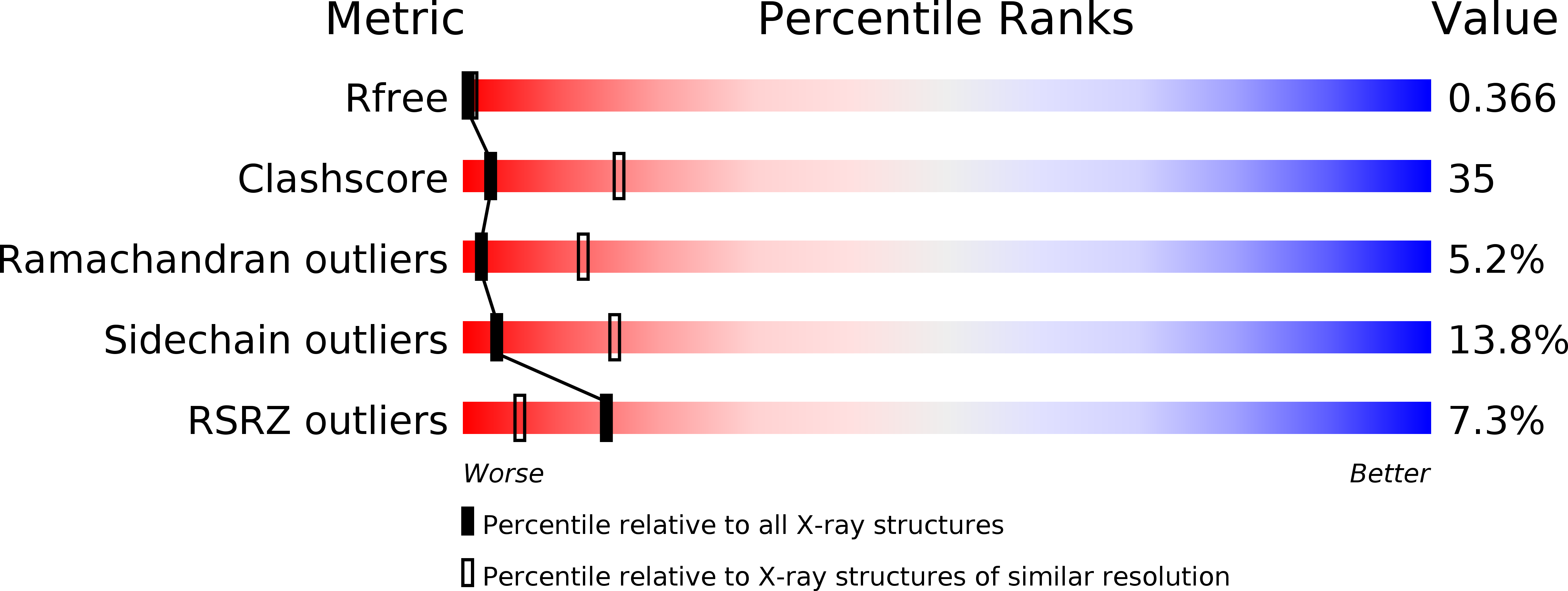
3.10 Å
R-Value Free:
0.35
R-Value Work:
0.31
R-Value Observed:
0.31
Space Group:
C 2 2 21
Macromolecular Entities
Structures with similar UniProt ID
Protein Blast
Polymer Type:polypeptide(L)
Molecule:Vinculin
Gene (Uniprot):VCL
Chain IDs:A
Chain Length:1069
Number of Molecules:1
Biological Source:Gallus gallus
Ligand Molecules
Primary Citation
Structural basis for vinculin activation at sites of cell adhesion.
Nature 430 583 586 (2004)
PMID: 15195105 DOI: 10.1038/nature02610

Abstact

Vinculin is a highly conserved intracellular protein with a crucial role in the maintenance and regulation of cell adhesion and migration. In the cytosol, vinculin adopts a default autoinhibited conformation. On recruitment to cell-cell and cell-matrix adherens-type junctions, vinculin becomes activated and mediates various protein-protein interactions that regulate the links between F-actin and the cadherin and integrin families of cell-adhesion molecules. Here we describe the crystal structure of the full-length vinculin molecule (1,066 amino acids), which shows a five-domain autoinhibited conformation in which the carboxy-terminal tail domain is held pincer-like by the vinculin head, and ligand binding is regulated both sterically and allosterically. We show that conformational changes in the head, tail and proline-rich domains are linked structurally and thermodynamically, and propose a combinatorial pathway to activation that ensures that vinculin is activated only at sites of cell adhesion when two or more of its binding partners are brought into apposition.

Legend

Protein

Chemical

Disease

Primary Citation of related structures