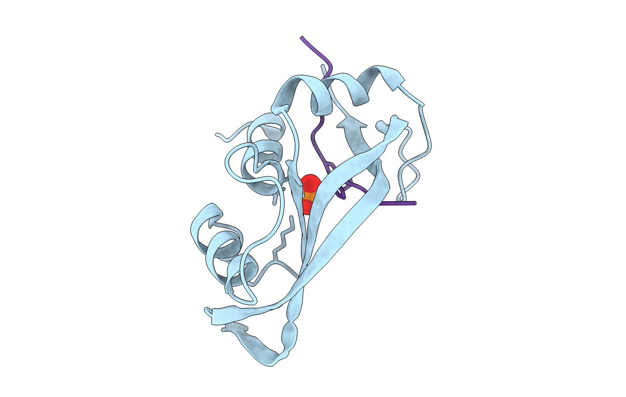
1SSC image
Deposition Date 1994-10-05
Release Date 1995-01-26
Last Version Date 2024-11-06
Entry Detail
PDB ID:
1SSC
Keywords:
Title:
THE 1.6 ANGSTROMS STRUCTURE OF A SEMISYNTHETIC RIBONUCLEASE CRYSTALLIZED FROM AQUEOUS ETHANOL. COMPARISON WITH CRYSTALS FROM SALT SOLUTIONS AND WITH RNASE A FROM AQUEOUS ALCOHOL SOLUTIONS
Biological Source:
Source Organism:
Bos taurus (Taxon ID: 9913)
Method Details:
Experimental Method:
Resolution:
2.00 Å
R-Value Observed:
0.16
Space Group:
P 1 21 1
Macromolecular Entities
Structures with similar UniProt ID
Protein Blast
Polymer Type:polypeptide(L)
Molecule:RIBONUCLEASE A
Gene (Uniprot):RNASE1
Chain IDs:A
Chain Length:112
Number of Molecules:1
Biological Source:Bos taurus
Structures with similar UniProt ID
Protein Blast
Polymer Type:polypeptide(L)
Molecule:RIBONUCLEASE A
Gene (Uniprot):RNASE1
Chain IDs:B
Chain Length:11
Number of Molecules:1
Biological Source:Bos taurus
Ligand Molecules
Primary Citation
1.6 A structure of semisynthetic ribonuclease crystallized from aqueous ethanol. Comparison with crystals from salt solutions and with ribonuclease A from aqueous alcohol solutions.
Acta Crystallogr.,Sect.D 51 1003 1012 (1995)
PMID: 15299768 DOI: 10.1107/S0907444995004574

Abstact

The non-covalent combination of residues 1-118 of RNase A with a synthetic 14-residue peptide containing residues 111-124 of the molecule forms a highly active semisynthetic enzyme, RNase 1-118:111-124. With this enzyme, the roles played by the six C-terminal residues in generating the catalytic efficiency and substrate specificity of RNase can be studied using chemically synthesized analogs. The structure of RNase 1-118:111-124 from 43% aqueous ethanol has been determined using molecular-replacement methods and refined to a crystallographic R-factor of 0.166 for all observed reflections in the range 7.0-1.6 A (Protein Data Bank file ISSC). The structure is compared with the 2.0 A structure of RNase A from 43% aqueous 2-methyl-2-propanol and with the 1.8 A structure of the semisynthetic enzyme obtained from crystals grown in concentrated salt solution. The structure of RNase 1-118:111-124 from aqueous ethanol is virtually identical to that of RNase A from aqueous 2-methyl-2-propanol. Half of the crystallographically bound water molecules are not coincident, however. The structure is somewhat less similar to that of RNase 1-118:111-124 from salt solutions, with a major difference being the positioning of active-site residue His119.

Legend

Protein

Chemical

Disease

Primary Citation of related structures