1SS9 image
Entry Detail
PDB ID:
1SS9
Keywords:
Title:
Crystal Structural Analysis of Active Site Mutant Q189E of LgtC
Biological Source:
Source Organism:
Host Organism:
PDB Version:
Deposition Date:
2004-03-23
Release Date:
2004-09-21
Method Details:
Experimental Method:
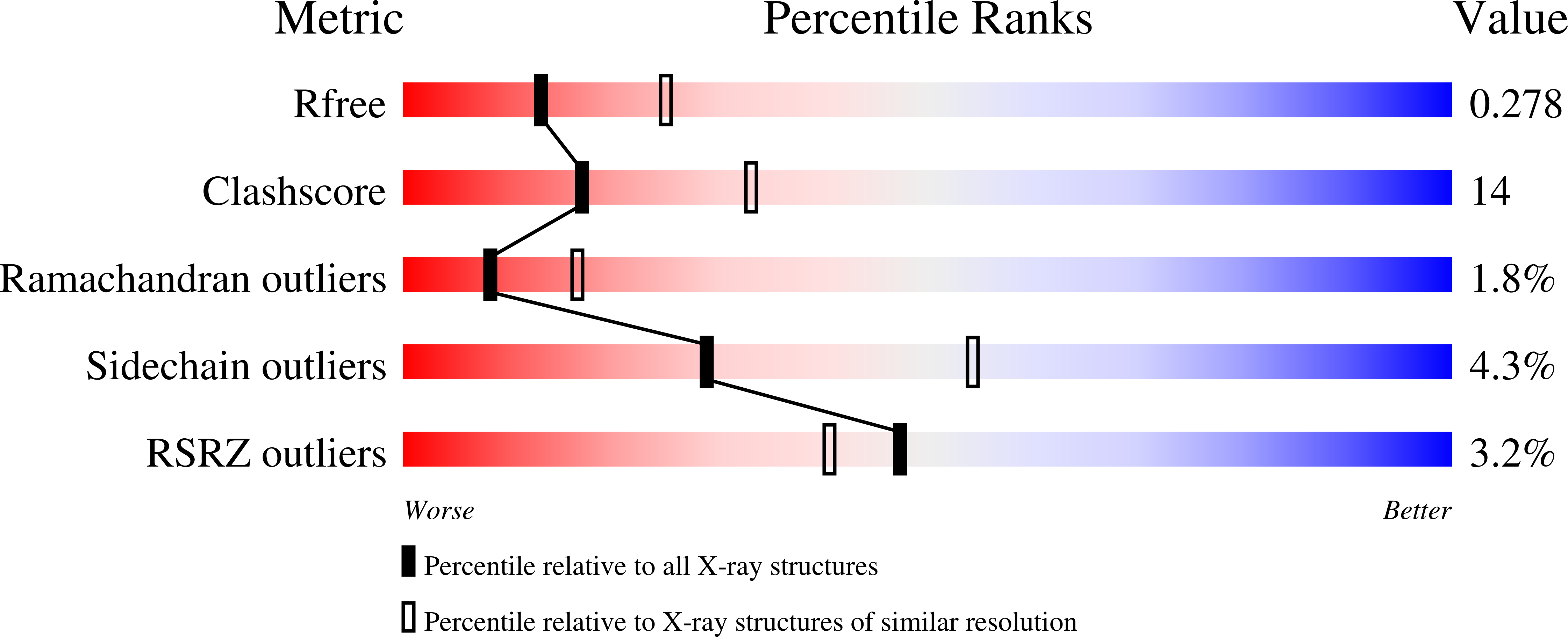
Resolution:
2.60 Å
R-Value Free:
0.28
R-Value Work:
0.21
R-Value Observed:
0.21
Space Group:
P 21 21 21
Macromolecular Entities
Polymer Type:polypeptide(L)
Description:alpha-1,4-galactosyl transferase
Mutations:Q189E
Chain IDs:A
Chain Length:311
Number of Molecules:1
Biological Source:Neisseria meningitidis
Primary Citation
Intermediate trapping on a mutant retaining alpha-galactosyltransferase identifies an unexpected aspartate residue.
J.Biol.Chem. 279 28339 28344 (2004)
PMID: 15075344 DOI: 10.1074/jbc.M400451200

Abstact

Lipopolysaccharyl-alpha-1,4-galactosyltransferase C (LgtC), a glycosyltransferase family 8 alpha-1,4-galactosyltransferase from Neisseria meningitidis, catalyzes the transfer of galactose from UDP galactose to terminal lactose-containing acceptor sugars with net retention of anomeric configuration. To investigate the potential role of discrete nucleophilic catalysis suggested by the double displacement mechanism generally proposed for retaining glycosyltransferases, the side chain amide of Gln-189, which is suitably positioned to act as the catalytic nucleophile of LgtC, was substituted with the more nucleophilic carboxylate-containing side chain of glutamate in the hope of accumulating a glycosyl-enzyme intermediate. The resulting mutant was subjected to kinetic, mass spectrometric, and x-ray crystallographic analysis. Although the K(m) for UDP-galactose is not significantly altered, the k(cat) was reduced to 3% that of the wild type enzyme. Electrospray mass spectrometric analysis revealed that a steady state population of the Q189E variant contains a covalently bound galactosyl moiety. Liquid chromatographic/mass spectrometric analysis of fragmented proteolytic digests identified the site of labeling not as Glu-189 but, surprisingly, as the sequentially adjacent Asp-190. However, the side chain carboxylate of Asp-190 is located 8.9 A away from the donor substrate in the available crystal structure. Kinetic analysis of a D190N mutant at this position revealed a k(cat) value 3000-fold lower than that of the wild type enzyme. A 2.6-A crystal structure of the Q189E mutant with bound uridine 5'-diphospho-2-deoxy-2-fluoro-alpha-d-galactopyranose revealed no significant perturbation of the mode of donor sugar binding nor of active site configuration. This is the first trapping of an intermediate in the active site of a retaining glycosyltransferase and, although not conclusive, implicates Asp-190 as an alternative candidate catalytic nucleophile, thereby rekindling a longstanding mechanistic debate.

Legend

Protein

Chemical

Disease

Primary Citation of related structures